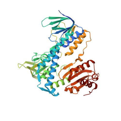
7ZYT

Crystal structure of the I318T pathogenic variant of the human dihydrolipoamide dehydrogenase


Experimental Data Snapshot

  • Method: X-RAY DIFFRACTION
  • Resolution: 2.89 Å
  • R-Value Free: 0.287 
  • R-Value Work: 0.250 
  • R-Value Observed: 0.252 

wwPDB Validation   3D Report Full Report


Ligand Structure Quality Assessment 


This is version 1.2 of the entry. See complete history


Literature

Structural and Biochemical Investigation of Selected Pathogenic Mutants of the Human Dihydrolipoamide Dehydrogenase.

Szabo, E.Nemes-Nikodem, E.Vass, K.R.Zambo, Z.Zrupko, E.Torocsik, B.Ozohanics, O.Nagy, B.Ambrus, A.

(2023) Int J Mol Sci 24

  • DOI: https://doi.org/10.3390/ijms241310826
  • Primary Citation of Related Structures:  
    7PSC, 7ZYT

  • PubMed Abstract: 

    Clinically relevant disease-causing variants of the human dihydrolipoamide dehydrogenase (hLADH, hE3), a common component of the mitochondrial α-keto acid dehydrogenase complexes, were characterized using a multipronged approach to unravel the molecular pathomechanisms that underlie hLADH deficiency. The G101del and M326V substitutions both reduced the protein stability and triggered the disassembly of the functional/obligate hLADH homodimer and significant FAD losses, which altogether eventually manifested in a virtually undetectable catalytic activity in both cases. The I12T-hLADH variant proved also to be quite unstable, but managed to retain the dimeric enzyme form; the LADH activity, both in the forward and reverse catalytic directions and the affinity for the prosthetic group FAD were both significantly compromised. None of the above three variants lent themselves to an in-depth structural analysis via X-ray crystallography due to inherent protein instability. Crystal structures at 2.89 and 2.44 Å resolutions were determined for the I318T- and I358T-hLADH variants, respectively; structure analysis revealed minor conformational perturbations, which correlated well with the residual LADH activities, in both cases. For the dimer interface variants G426E-, I445M-, and R447G-hLADH, enzyme activities and FAD loss were determined and compared against the previously published structural data.


  • Organizational Affiliation

    Department of Biochemistry, Institute of Biochemistry and Molecular Biology, Semmelweis University, 37-47 Tuzolto St., 1094 Budapest, Hungary.


Macromolecules
Find similar proteins by:  (by identity cutoff)  |  3D Structure
Entity ID: 1
MoleculeChains Sequence LengthOrganismDetailsImage
Dihydrolipoyl dehydrogenase, mitochondrial
A, B
496Homo sapiensMutation(s): 1 
Gene Names: DLDGCSLLADPHE3
EC: 1.8.1.4
UniProt & NIH Common Fund Data Resources
Find proteins for P09622 (Homo sapiens)
Explore P09622 
Go to UniProtKB:  P09622
PHAROS:  P09622
GTEx:  ENSG00000091140 
Entity Groups  
Sequence Clusters30% Identity50% Identity70% Identity90% Identity95% Identity100% Identity
UniProt GroupP09622
Sequence Annotations
Expand
  • Reference Sequence
Experimental Data & Validation

Experimental Data

  • Method: X-RAY DIFFRACTION
  • Resolution: 2.89 Å
  • R-Value Free: 0.287 
  • R-Value Work: 0.250 
  • R-Value Observed: 0.252 
  • Space Group: C 1 2 1
Unit Cell:
Length ( Å )Angle ( ˚ )
a = 188.65α = 90
b = 59β = 101.4
c = 83.23γ = 90
Software Package:
Software NamePurpose
MxCuBEdata collection
XDSdata reduction
XSCALEdata scaling
MOLREPphasing
PHENIXrefinement
PDB_EXTRACTdata extraction

Structure Validation

View Full Validation Report


Ligand Structure Quality Assessment 


Entry History & Funding Information

Deposition Data


Funding OrganizationLocationGrant Number
Hungarian Academy of SciencesHungaryKTIA_13_NAP_III/6 and 2017-1.2.1-NKP-2017-00002 [to A.-V.V.]
European Union (EU)European UnionHorizon 2020 Research and Innovation Programme MX-212-00269 ST [to A.A.]
Hungarian National Research, Development and Innovation OfficeHungaryTKP2021-EGA-25

Revision History  (Full details and data files)

  • Version 1.0: 2023-06-14
    Type: Initial release
  • Version 1.1: 2023-07-26
    Changes: Database references
  • Version 1.2: 2024-02-07
    Changes: Data collection, Refinement description