The structure of a Bacteroides thetaiotaomicron carbohydrate-binding module provides new insight into the recognition of complex pectic polysaccharides by the human microbiome.
Trovao, F., Correia, V.G., Lourenco, F.M., Ribeiro, D.O., Carvalho, A.L., Palma, A.S., Pinheiro, B.A.(2023) J Struct Biol X 7: 100084-100084
- PubMed: 36660365 Search on PubMedSearch on PubMed Central
- DOI: https://doi.org/10.1016/j.yjsbx.2022.100084
- Primary Citation Related Structures:
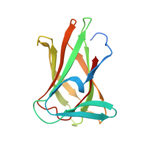
7ZVO - PubMed Abstract:
The Bacteroides thetaiotaomicron has developed a consortium of enzymes capable of overcoming steric constraints and degrading, in a sequential manner, the complex rhamnogalacturonan II (RG-II) polysaccharide. BT0996 protein acts in the initial stages of the RG-II depolymerisation, where its two catalytic modules remove the terminal monosaccharides from RG-II side chains A and B. BT0996 is modular and has three putative carbohydrate-binding modules (CBMs) for which the roles in the RG-II degradation are unknown. Here, we present the characterisation of the module at the C-terminal domain, which we designated BT0996-C. The high-resolution structure obtained by X-ray crystallography reveals that the protein displays a typical β-sandwich fold with structural similarity to CBMs assigned to families 6 and 35. The distinctive features are: 1) the presence of several charged residues at the BT0996-C surface creating a large, broad positive lysine-rich patch that encompasses the putative binding site; and 2) the absence of the highly conserved binding-site signatures observed in CBMs from families 6 and 35, such as region A tryptophan and region C asparagine. These findings hint at a binding mode of BT0996-C not yet observed in its homologues. In line with this, carbohydrate microarrays and microscale thermophoresis show the ability of BT0996-C to bind α1-4-linked polygalacturonic acid, and that electrostatic interactions are essential for the recognition of the anionic polysaccharide. The results support the hypothesis that BT0996-C may have evolved to potentiate the action of BT0996 catalytic modules on the complex structure of RG-II by binding to the polygalacturonic acid backbone sequence.
- UCIBIO - Applied Molecular Biosciences Unit, Department of Chemistry, NOVA School of Science and Technology, Universidade NOVA de Lisboa, 2829-516 Caparica, Portugal.
Organizational Affiliation: