7ZUI

PENICILLIN-BINDING PROTEIN 1B (PBP-1B) in complex with lactone 5Az - Streptococcus pneumoniae R6


Experimental Data Snapshot

  • Method: X-RAY DIFFRACTION
  • Resolution: 1.57 Å
  • R-Value Free: 0.181 
  • R-Value Work: 0.154 

wwPDB Validation   3D Report Full Report


Ligand Structure Quality Assessment 


This is version 1.2 of the entry. See complete history

Re-refinement Note

A newer entry is available that reflects an alternative modeling of the original data: 7ZUH 7ZUJ 7ZUK 7ZUL


Literature

Combined Structural Analysis and Molecular Dynamics Reveal Penicillin-Binding Protein Inhibition Mode with beta-Lactones.

Flanders, P.L.Contreras-Martel, C.Brown, N.W.Shirley, J.D.Martins, A.Nauta, K.N.Dessen, A.Carlson, E.E.Ambrose, E.A.

(2022) ACS Chem Biol 17: 3110-3120

  • DOI: https://doi.org/10.1021/acschembio.2c00503
  • Primary Citation of Related Structures:  
    7ZUH, 7ZUI, 7ZUJ, 7ZUK, 7ZUL

  • PubMed Abstract: 

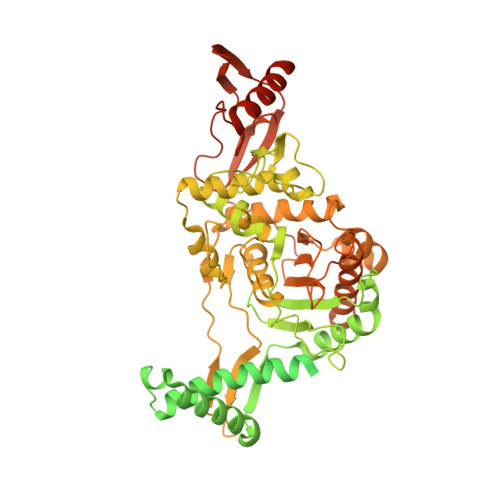
    β-Lactam antibiotics comprise one of the most widely used therapeutic classes to combat bacterial infections. This general scaffold has long been known to inhibit bacterial cell wall biosynthesis by inactivating penicillin-binding proteins (PBPs); however, bacterial resistance to β-lactams is now widespread, and new strategies are urgently needed to target PBPs and other proteins involved in bacterial cell wall formation. A key requirement in the identification of strategies to overcome resistance is a deeper understanding of the roles of the PBPs and their associated proteins during cell growth and division, such as can be obtained with the use of selective chemical probes. Probe development has typically depended upon known PBP inhibitors, which have historically been thought to require a negatively charged moiety that mimics the C-terminus of the PBP natural peptidoglycan substrate, d-Ala-d-Ala. However, we have identified a new class of β-lactone-containing molecules that interact with PBPs, often in an isoform-specific manner, and do not incorporate this C-terminal mimetic. Here, we report a series of structural biology experiments and molecular dynamics simulations that we utilized to evaluate specific binding modes of this novel PBP inhibitor class. In this work, we obtained <2 Å resolution X-ray structures of four β-lactone probes bound to PBP1b from Streptococcus pneumoniae . Despite their diverging recognition modes beyond the site of covalent modification, these four probes all efficiently labeled PBP1b, as well as other PBPs from S . pneumoniae . From these structures, we analyzed protein-ligand interactions and characterized the β-lactone-bound active sites using in silico mutagenesis and molecular dynamics. Our approach has clarified the dynamic interaction profile in this series of ligands, expanding the understanding of PBP inhibitor binding.


  • Organizational Affiliation

    Department of Medicinal Chemistry, University of Minnesota, 208 Harvard Street SE, Minneapolis, Minnesota 55454, United States.


Macromolecules
Find similar proteins by:  (by identity cutoff)  |  3D Structure
Entity ID: 1
MoleculeChains Sequence LengthOrganismDetailsImage
Penicillin-binding protein 1bA [auth AAA]821Streptococcus pneumoniae R6Mutation(s): 3 
Gene Names: pbp1bspr1909
EC: 2.3.2 (PDB Primary Data), 2.4.1.129 (PDB Primary Data)
UniProt
Find proteins for Q7CRA4 (Streptococcus pneumoniae (strain ATCC BAA-255 / R6))
Explore Q7CRA4 
Go to UniProtKB:  Q7CRA4
Entity Groups  
Sequence Clusters30% Identity50% Identity70% Identity90% Identity95% Identity100% Identity
UniProt GroupQ7CRA4
Sequence Annotations
Expand
  • Reference Sequence
Small Molecules
Ligands 3 Unique
IDChains Name / Formula / InChI Key2D Diagram3D Interactions
K2O (Subject of Investigation/LOI)
Query on K2O

Download Ideal Coordinates CCD File 
B [auth AAA]6-azido-N-[(2R)-1-oxidanylidene-1-[[(2S,3R)-3-oxidanyl-1-oxidanylidene-butan-2-yl]amino]propan-2-yl]hexanamide
C13 H23 N5 O4
QSZPURBQGQCCCA-GMTAPVOTSA-N
DMS
Query on DMS

Download Ideal Coordinates CCD File 
BA [auth AAA]DIMETHYL SULFOXIDE
C2 H6 O S
IAZDPXIOMUYVGZ-UHFFFAOYSA-N
CL
Query on CL

Download Ideal Coordinates CCD File 
AA [auth AAA]
C [auth AAA]
D [auth AAA]
E [auth AAA]
F [auth AAA]
AA [auth AAA],
C [auth AAA],
D [auth AAA],
E [auth AAA],
F [auth AAA],
G [auth AAA],
H [auth AAA],
I [auth AAA],
J [auth AAA],
K [auth AAA],
L [auth AAA],
M [auth AAA],
N [auth AAA],
O [auth AAA],
P [auth AAA],
Q [auth AAA],
R [auth AAA],
S [auth AAA],
T [auth AAA],
U [auth AAA],
V [auth AAA],
W [auth AAA],
X [auth AAA],
Y [auth AAA],
Z [auth AAA]
CHLORIDE ION
Cl
VEXZGXHMUGYJMC-UHFFFAOYSA-M
Experimental Data & Validation

Experimental Data

  • Method: X-RAY DIFFRACTION
  • Resolution: 1.57 Å
  • R-Value Free: 0.181 
  • R-Value Work: 0.154 
  • Space Group: C 2 2 21
Unit Cell:
Length ( Å )Angle ( ˚ )
a = 96.025α = 90
b = 149.061β = 90
c = 99.226γ = 90
Software Package:
Software NamePurpose
REFMACrefinement
XDSdata reduction
XSCALEdata scaling
PHASERphasing

Structure Validation

View Full Validation Report


Ligand Structure Quality Assessment 


Entry History & Funding Information

Deposition Data


Funding OrganizationLocationGrant Number
Agence Nationale de la Recherche (ANR)FranceANR-10-INBS-0005-02
Agence Nationale de la Recherche (ANR)FranceANR-17-EURE-0003
National Institutes of Health/National Center for Research Resources (NIH/NCRR)United StatesR01 GM128439
National Institutes of Health/National Center for Research Resources (NIH/NCRR)United StatesK12 GM119955
National Institutes of Health/National Center for Research Resources (NIH/NCRR)United StatesTL1R002493
National Institutes of Health/National Center for Research Resources (NIH/NCRR)United StatesUL1TR002494

Revision History  (Full details and data files)

  • Version 1.0: 2022-11-02
    Type: Initial release
  • Version 1.1: 2022-11-30
    Changes: Database references
  • Version 1.2: 2024-01-31
    Changes: Data collection, Refinement description