7ZUD | pdb_00007zud

Crystal structure of HIV-1 capsid IP6-CPSF6 complex


Experimental Data Snapshot

  • Method: X-RAY DIFFRACTION
  • Resolution: 2.93 Å
  • R-Value Free: 
    0.336 (Depositor), 0.341 (DCC) 
  • R-Value Work: 
    0.278 (Depositor), 0.279 (DCC) 
  • R-Value Observed: 
    0.278 (Depositor) 

Starting Model: experimental
View more details

wwPDB Validation 3D Report Full Report

Validation slider image for 7ZUD

Ligand Structure Quality Assessment 


This is version 1.2 of the entry. See complete history

Literature

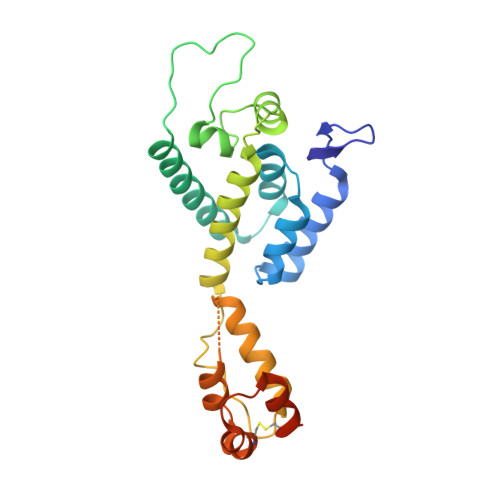
CP-MAS and Solution NMR Studies of Allosteric Communication in CA-assemblies of HIV-1.

Nicastro, G.Lucci, M.Oregioni, A.Kelly, G.Frenkiel, T.A.Taylor, I.A.

(2022) J Mol Biology 434: 167691-167691

  • DOI: https://doi.org/10.1016/j.jmb.2022.167691
  • Primary Citation Related Structures: 
    7ZUD

  • PubMed Abstract: 

    Solution and solid-state NMR spectroscopy are highly complementary techniques for studying structure and dynamics in very high molecular weight systems. Here we have analysed the dynamics of HIV-1 capsid (CA) assemblies in presence of the cofactors IP6 and ATPγS and the host-factor CPSF6 using a combination of solution state and cross polarisation magic angle spinning (CP-MAS) solid-state NMR. In particular, dynamical effects on ns to µs and µs to ms timescales are observed revealing diverse motions in assembled CA. Using CP-MAS NMR, we exploited the sensitivity of the amide/Cα-Cβ backbone chemical shifts in DARR and NCA spectra to observe the plasticity of the HIV-1 CA tubular assemblies and also map the binding of cofactors and the dynamics of cofactor-CA complexes. In solution, we measured how the addition of host- and co-factors to CA -hexamers perturbed the chemical shifts and relaxation properties of CA-Ile and -Met methyl groups using transverse-relaxation-optimized NMR spectroscopy to exploit the sensitivity of methyl groups as probes in high-molecular weight proteins. These data show how dynamics of the CA protein assembly over a range of spatial and temporal scales play a critical role in CA function. Moreover, we show that binding of IP6, ATPγS and CPSF6 results in local chemical shift as well as dynamic changes for a significant, contiguous portion of CA, highlighting how allosteric pathways communicate ligand interactions between adjacent CA protomers.


  • Organizational Affiliation
    • Macromolecular Structure Laboratory, The Francis Crick Institute, 1 Midland Road, London NW1 1AT, UK.

Macromolecule Content 

  • Total Structure Weight: 28.4 kDa 
  • Atom Count: 1,849 
  • Modeled Residue Count: 223 
  • Deposited Residue Count: 246 
  • Unique protein chains: 2

Macromolecules

Find similar proteins by:|  3D Structure
Entity ID: 1
MoleculeChains  Sequence LengthOrganismDetailsImage
Capsid protein p24233Human immunodeficiency virus 1Mutation(s): 4 
Gene Names: gag-pol
UniProt
Find proteins for P12497 (Human immunodeficiency virus type 1 group M subtype B (isolate NY5))
Explore P12497 
Go to UniProtKB:  P12497
Entity Groups
Sequence Clusters30% Identity50% Identity70% Identity90% Identity95% Identity100% Identity
UniProt GroupP12497
Sequence Annotations
Expand
Reference Sequence
Find similar proteins by:  Sequence   |   3D Structure  
Entity ID: 2
MoleculeChains  Sequence LengthOrganismDetailsImage
Cleavage and polyadenylation specificity factor subunit 6B [auth M]13Homo sapiensMutation(s): 0 
UniProt & NIH Common Fund Data Resources
Find proteins for Q16630 (Homo sapiens)
Explore Q16630 
Go to UniProtKB:  Q16630
PHAROS:  Q16630
GTEx:  ENSG00000111605 
Entity Groups
Sequence Clusters30% Identity50% Identity70% Identity90% Identity95% Identity100% Identity
UniProt GroupQ16630
Sequence Annotations
Expand
Reference Sequence

Experimental Data & Validation

Experimental Data

  • Method: X-RAY DIFFRACTION
  • Resolution: 2.93 Å
  • R-Value Free:  0.336 (Depositor), 0.341 (DCC) 
  • R-Value Work:  0.278 (Depositor), 0.279 (DCC) 
  • R-Value Observed: 0.278 (Depositor) 
Space Group: P 6
Unit Cell:
Length ( Å )Angle ( ˚ )
a = 90.741α = 90
b = 90.741β = 90
c = 56.588γ = 120
Software Package:
Software NamePurpose
PHENIXrefinement
autoPROCdata processing
STARANISOdata scaling
autoPROCdata reduction
PHASERphasing

Structure Validation

View Full Validation Report


Ligand Structure Quality Assessment 


Entry History 

& Funding Information

Deposition Data


Funding OrganizationLocationGrant Number
The Francis Crick InstituteUnited KingdomFC001178
Wellcome TrustUnited Kingdom108014/Z/15/Z

Revision History  (Full details and data files)

  • Version 1.0: 2022-07-27
    Type: Initial release
  • Version 1.1: 2024-01-31
    Changes: Data collection, Refinement description
  • Version 1.2: 2024-10-23
    Changes: Structure summary