7ZR1 | pdb_00007zr1

Chaetomium thermophilum Mre11-Rad50-Nbs1 complex bound to ATPyS (composite structure)


Experimental Data Snapshot

  • Method: ELECTRON MICROSCOPY
  • Resolution: 4.00 Å
  • Aggregation State: PARTICLE 
  • Reconstruction Method: SINGLE PARTICLE 

wwPDB Validation 3D Report Full Report

Validation slider image for 7ZR1

This is version 1.2 of the entry. See complete history

Literature

Cryo-EM structure of the Mre11-Rad50-Nbs1 complex reveals the molecular mechanism of scaffolding functions.

Rotheneder, M.Stakyte, K.van de Logt, E.Bartho, J.D.Lammens, K.Fan, Y.Alt, A.Kessler, B.Jung, C.Roos, W.P.Steigenberger, B.Hopfner, K.P.

(2023) Mol Cell 83: 167-185.e9

  • DOI: https://doi.org/10.1016/j.molcel.2022.12.003
  • Primary Citation Related Structures: 
    7ZQY, 7ZR1, 8BAH

  • PubMed Abstract: 

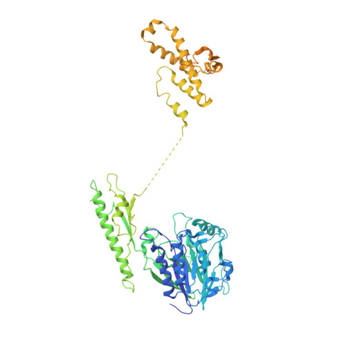
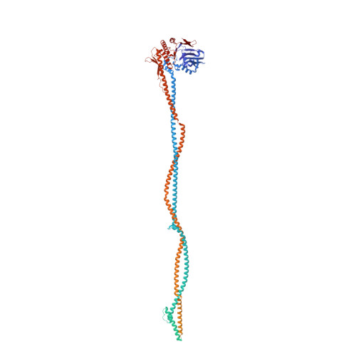
    The DNA double-strand break repair complex Mre11-Rad50-Nbs1 (MRN) detects and nucleolytically processes DNA ends, activates the ATM kinase, and tethers DNA at break sites. How MRN can act both as nuclease and scaffold protein is not well understood. The cryo-EM structure of MRN from Chaetomium thermophilum reveals a 2:2:1 complex with a single Nbs1 wrapping around the autoinhibited Mre11 nuclease dimer. MRN has two DNA-binding modes, one ATP-dependent mode for loading onto DNA ends and one ATP-independent mode through Mre11's C terminus, suggesting how it may interact with DSBs and intact DNA. MRNs two 60-nm-long coiled-coil domains form a linear rod structure, the apex of which is assembled by the two joined zinc-hook motifs. Apices from two MRN complexes can further dimerize, forming 120-nm spanning MRN-MRN structures. Our results illustrate the architecture of MRN and suggest how it mechanistically integrates catalytic and tethering functions.


  • Organizational Affiliation
    • Gene Center, Department of Biochemistry, Ludwig Maximilians Universität, Munich, Germany.

Macromolecule Content 

  • Total Structure Weight: 577.83 kDa 
  • Atom Count: 21,793 
  • Modeled Residue Count: 2,686 
  • Deposited Residue Count: 5,044 
  • Unique protein chains: 3

Macromolecules

Find similar proteins by:|  3D Structure
Entity ID: 1
MoleculeChains  Sequence LengthOrganismDetailsImage
Double-strand break repair protein
A, B
730Thermochaetoides thermophila DSM 1495Mutation(s): 0 
Gene Names: CTHT_0007600
EC: 3.1
UniProt
Find proteins for G0RYR3 (Chaetomium thermophilum (strain DSM 1495 / CBS 144.50 / IMI 039719))
Explore G0RYR3 
Go to UniProtKB:  G0RYR3
Entity Groups
Sequence Clusters30% Identity50% Identity70% Identity90% Identity95% Identity100% Identity
UniProt GroupG0RYR3
Sequence Annotations
Expand
Reference Sequence
Find similar proteins by:|  3D Structure
Entity ID: 2
MoleculeChains  Sequence LengthOrganismDetailsImage
DH domain-containing protein
C, D
1,315Thermochaetoides thermophilaMutation(s): 0 
Gene Names: CTHT_0073630
EC: 3.6
UniProt
Find proteins for G0SHW7 (Chaetomium thermophilum (strain DSM 1495 / CBS 144.50 / IMI 039719))
Explore G0SHW7 
Go to UniProtKB:  G0SHW7
Entity Groups
Sequence Clusters30% Identity50% Identity70% Identity90% Identity95% Identity100% Identity
UniProt GroupG0SHW7
Sequence Annotations
Expand
Reference Sequence
Find similar proteins by:|  3D Structure
Entity ID: 3
MoleculeChains  Sequence LengthOrganismDetailsImage
FHA domain-containing protein954Thermochaetoides thermophila DSM 1495Mutation(s): 0 
Gene Names: CTHT_0047880
UniProt
Find proteins for G0SAV1 (Chaetomium thermophilum (strain DSM 1495 / CBS 144.50 / IMI 039719))
Explore G0SAV1 
Go to UniProtKB:  G0SAV1
Entity Groups
Sequence Clusters30% Identity50% Identity70% Identity90% Identity95% Identity100% Identity
UniProt GroupG0SAV1
Sequence Annotations
Expand
Reference Sequence

Small Molecules

Ligands 3 Unique
IDChains Name / Formula / InChI Key2D Diagram3D Interactions
AGS
(Subject of Investigation/LOI)

Query on AGS


Download:Ideal Coordinates CCD File
K [auth C],
M [auth D]
PHOSPHOTHIOPHOSPHORIC ACID-ADENYLATE ESTER
C10 H16 N5 O12 P3 S
NLTUCYMLOPLUHL-KQYNXXCUSA-N
MN
(Subject of Investigation/LOI)

Query on MN


Download:Ideal Coordinates CCD File
F [auth A],
G [auth A],
H [auth B],
I [auth B]
MANGANESE (II) ION
Mn
WAEMQWOKJMHJLA-UHFFFAOYSA-N
MG
(Subject of Investigation/LOI)

Query on MG


Download:Ideal Coordinates CCD File
J [auth C],
L [auth D]
MAGNESIUM ION
Mg
JLVVSXFLKOJNIY-UHFFFAOYSA-N

Experimental Data & Validation

Experimental Data

  • Method: ELECTRON MICROSCOPY
  • Resolution: 4.00 Å
  • Aggregation State: PARTICLE 
  • Reconstruction Method: SINGLE PARTICLE 

Structure Validation

View Full Validation Report


Entry History 

& Funding Information

Deposition Data


Funding OrganizationLocationGrant Number
German Research Foundation (DFG)GermanyCRC1361
German Research Foundation (DFG)GermanySFB1361
German Research Foundation (DFG)GermanyGRK1721

Revision History  (Full details and data files)

  • Version 1.0: 2023-01-11
    Type: Initial release
  • Version 1.1: 2023-02-01
    Changes: Database references
  • Version 1.2: 2023-12-13
    Changes: Data collection