7ZP0

Crystal structure of CusS histidine kinase catalytic core from Escherichia coli


Experimental Data Snapshot

  • Method: X-RAY DIFFRACTION
  • Resolution: 1.40 Å
  • R-Value Free: 0.219 
  • R-Value Work: 0.177 
  • R-Value Observed: 0.177 

wwPDB Validation   3D Report Full Report


This is version 1.2 of the entry. See complete history


Literature

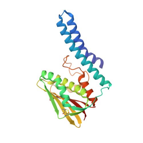
Crystal structure of the Escherichia coli CusS kinase core.

Cociurovscaia, A.Bujacz, G.Pietrzyk-Brzezinska, A.J.

(2022) J Struct Biol 214: 107883-107883

  • DOI: https://doi.org/10.1016/j.jsb.2022.107883
  • Primary Citation of Related Structures:  
    7ZP0

  • PubMed Abstract: 

    The CusS histidine kinase is a member of Escherichia coli two-component signal transduction system, engaged in a response to copper ions excess in the cell periplasm. The periplasmic sensor domain of CusS binds the free copper ions and the CusS kinase core phosphorylates the cognate CusR which regulates transcription of the efflux pomp CusCBA. A small amount of copper ions is indispensable for the aerobic cell metabolism. Nonetheless, its excess in the cytoplasm generates damaging and reactive hydroxyl radicals. For that reason, understanding the bacterial copper sensing mechanisms can contribute to reducing bacterial copper-resistance and developing bactericidal copper-based materials. The crystal structure of the CusS kinase core was solved at the resolution of 1.4 Å. The cytoplasmic catalytic core domains formed a homodimer. Based on the obtained structure, the intramolecular and intermolecular interactions crucial for the mechanism of CusS autophosphorylation were described.


  • Organizational Affiliation

    Institute of Molecular and Industrial Biotechnology, Faculty of Biotechnology and Food Sciences, Lodz University of Technology, Lodz 90-537, Poland.


Macromolecules
Find similar proteins by:  (by identity cutoff)  |  3D Structure
Entity ID: 1
MoleculeChains Sequence LengthOrganismDetailsImage
Sensor protein
A, B, C, D, E
A, B, C, D, E, F
217Escherichia coli 'BL21-Gold(DE3)pLysS AGMutation(s): 1 
Gene Names: cusSCWS33_25405GNZ05_00640NCTC8985_02504
EC: 2.7.13.3
UniProt
Find proteins for A0A140NBW3 (Escherichia coli (strain B / BL21-DE3))
Explore A0A140NBW3 
Go to UniProtKB:  A0A140NBW3
Entity Groups  
Sequence Clusters30% Identity50% Identity70% Identity90% Identity95% Identity100% Identity
UniProt GroupA0A140NBW3
Sequence Annotations
Expand
  • Reference Sequence
Experimental Data & Validation

Experimental Data

  • Method: X-RAY DIFFRACTION
  • Resolution: 1.40 Å
  • R-Value Free: 0.219 
  • R-Value Work: 0.177 
  • R-Value Observed: 0.177 
  • Space Group: P 1 21 1
Unit Cell:
Length ( Å )Angle ( ˚ )
a = 69.757α = 90
b = 129.095β = 92.09
c = 69.834γ = 90
Software Package:
Software NamePurpose
PHENIXrefinement
XSCALEdata scaling
PDB_EXTRACTdata extraction
XDSdata reduction
PHENIXphasing

Structure Validation

View Full Validation Report


Entry History & Funding Information

Deposition Data


Funding OrganizationLocationGrant Number
Not funded--

Revision History  (Full details and data files)

  • Version 1.0: 2022-08-31
    Type: Initial release
  • Version 1.1: 2024-01-31
    Changes: Data collection, Refinement description
  • Version 1.2: 2024-04-10
    Changes: Structure summary