Pharmacological blocking of microfibrillar-associated protein 4 reduces retinal neoangiogenesis and vascular leakage.
Schlosser, A., Pilecki, B., Allen, C., Benest, A.V., Lynch, A.P., Hua, J., Ved, N., Blackley, Z., Andersen, T.L., Hennig, D., Graversen, J.H., Moller, S., Skallerup, S., Ormhoj, M., Lange, C., Agostini, H.T., Grauslund, J., Heegaard, S., Dacheva, I., Koss, M., Hu, W., Iglesias, B., Lawrence, M.S., Beck, H.C., Steffensen, L.B., Laursen, N.S., Andersen, G.R., Holmskov, U., Bates, D.O., Sorensen, G.L.(2025) Mol Ther
- PubMed: 39863929 Search on PubMed
- DOI: https://doi.org/10.1016/j.ymthe.2025.01.038
- Primary Citation Related Structures:
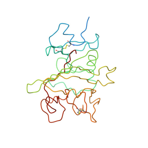
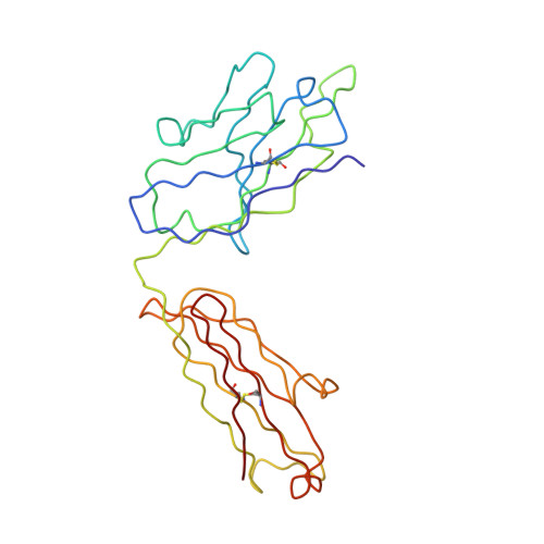
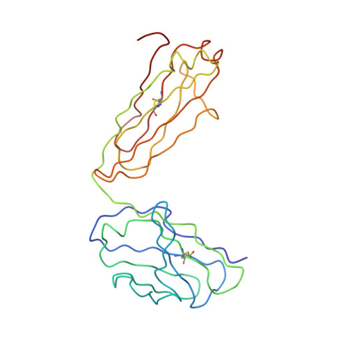
7ZMK - PubMed Abstract:
Neovascular age-related macular degeneration and diabetic macular edema are leading causes of vision-loss evoked by retinal neovascularization and vascular leakage. The glycoprotein microfibrillar-associated protein 4 (MFAP4) is an integrin α V β 3/5/6 ligand present in the extracellular matrix. Single-cell transcriptomics reveal MFAP4 expression in cell-types in close proximity to vascular endothelial cells including choroidal vascular mural cells and retinal astrocytes and Müller cells. Binding of the anti-MFAP4 antibody, hAS0326, makes MFAP4 inaccessible for integrin receptor interaction and thereby hAS0326 blocked endothelial cell motility in vitro. Intravitreal hAS0326 inhibited retinal vascular lesion area and neovessel volume in a laser-induced choroidal neovascularization mouse model, vascular permeability in streptozotocin-induced retinopathy and vascular leakage area in a chronic non-human primate model of DL-2-aminoadipic acid-induced retinopathy. One dose of hAS0326 showed duration of efficacy of at least 12 weeks in the latter model. Moreover, hAS0326-treatment significantly enriched gene ontology terms involving reduction of integrin binding. Our data suggest that hAS0326 constitutes a promising treatment of neovascularization and vascular leakage in retinal diseases.
- Department of Molecular Medicine, University of Southern Denmark; Odense, 5230, Denmark.
Organizational Affiliation: