7ZHQ

Crystal structure of TTBK1 in complex with compound 10 (7-009)


Experimental Data Snapshot

  • Method: X-RAY DIFFRACTION
  • Resolution: 1.80 Å
  • R-Value Free: 0.212 
  • R-Value Work: 0.174 
  • R-Value Observed: 0.176 

wwPDB Validation   3D Report Full Report


Ligand Structure Quality Assessment 


This is version 1.2 of the entry. See complete history


Literature

Modulation of tau tubulin kinases (TTBK1 and TTBK2) impacts ciliogenesis.

Bashore, F.M.Marquez, A.B.Chaikuad, A.Howell, S.Dunn, A.S.Beltran, A.A.Smith, J.L.Drewry, D.H.Beltran, A.S.Axtman, A.D.

(2023) Sci Rep 13: 6118-6118

  • DOI: https://doi.org/10.1038/s41598-023-32854-4
  • Primary Citation of Related Structures:  
    7ZHN, 7ZHO, 7ZHP, 7ZHQ

  • PubMed Abstract: 

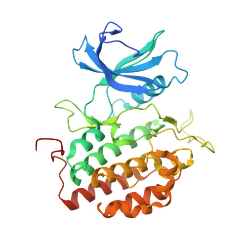
    Tau tubulin kinase 1 and 2 (TTBK1/2) are highly homologous kinases that are expressed and mediate disease-relevant pathways predominantly in the brain. Distinct roles for TTBK1 and TTBK2 have been delineated. While efforts have been devoted to characterizing the impact of TTBK1 inhibition in diseases like Alzheimer's disease and amyotrophic lateral sclerosis, TTBK2 inhibition has been less explored. TTBK2 serves a critical function during cilia assembly. Given the biological importance of these kinases, we designed a targeted library from which we identified several chemical tools that engage TTBK1 and TTBK2 in cells and inhibit their downstream signaling. Indolyl pyrimidinamine 10 significantly reduced the expression of primary cilia on the surface of human induced pluripotent stem cells (iPSCs). Furthermore, analog 10 phenocopies TTBK2 knockout in iPSCs, confirming a role for TTBK2 in ciliogenesis.


  • Organizational Affiliation

    Structural Genomics Consortium, UNC Eshelman School of Pharmacy, University of North Carolina at Chapel Hill, Chapel Hill, NC, 27599, USA.


Macromolecules
Find similar proteins by:  (by identity cutoff)  |  3D Structure
Entity ID: 1
MoleculeChains Sequence LengthOrganismDetailsImage
Tau-tubulin kinase 1309Homo sapiensMutation(s): 0 
Gene Names: TTBK1BDTKKIAA1855
EC: 2.7.11.1
UniProt & NIH Common Fund Data Resources
Find proteins for Q5TCY1 (Homo sapiens)
Explore Q5TCY1 
Go to UniProtKB:  Q5TCY1
PHAROS:  Q5TCY1
GTEx:  ENSG00000146216 
Entity Groups  
Sequence Clusters30% Identity50% Identity70% Identity90% Identity95% Identity100% Identity
UniProt GroupQ5TCY1
Sequence Annotations
Expand
  • Reference Sequence
Small Molecules
Ligands 3 Unique
IDChains Name / Formula / InChI Key2D Diagram3D Interactions
IRD (Subject of Investigation/LOI)
Query on IRD

Download Ideal Coordinates CCD File 
L [auth A](3~{S})-1-(4-azanyl-3,5,12-triazatetracyclo[9.7.0.0^{2,7}.0^{13,18}]octadeca-1(11),2,4,6,13(18),14,16-heptaen-16-yl)-3-methyl-pent-1-yn-3-ol
C21 H22 N4 O
ALQLTPNCVCECNJ-NRFANRHFSA-N
PO4
Query on PO4

Download Ideal Coordinates CCD File 
B [auth A]PHOSPHATE ION
O4 P
NBIIXXVUZAFLBC-UHFFFAOYSA-K
EDO
Query on EDO

Download Ideal Coordinates CCD File 
C [auth A]
D [auth A]
E [auth A]
F [auth A]
G [auth A]
C [auth A],
D [auth A],
E [auth A],
F [auth A],
G [auth A],
H [auth A],
I [auth A],
J [auth A],
K [auth A]
1,2-ETHANEDIOL
C2 H6 O2
LYCAIKOWRPUZTN-UHFFFAOYSA-N
Experimental Data & Validation

Experimental Data

  • Method: X-RAY DIFFRACTION
  • Resolution: 1.80 Å
  • R-Value Free: 0.212 
  • R-Value Work: 0.174 
  • R-Value Observed: 0.176 
  • Space Group: C 1 2 1
Unit Cell:
Length ( Å )Angle ( ˚ )
a = 171.308α = 90
b = 39.543β = 103.23
c = 49.65γ = 90
Software Package:
Software NamePurpose
REFMACrefinement
Aimlessdata scaling
PDB_EXTRACTdata extraction
XDSdata reduction
PHASERphasing

Structure Validation

View Full Validation Report


Ligand Structure Quality Assessment 


Entry History & Funding Information

Deposition Data


Funding OrganizationLocationGrant Number
Not funded--

Revision History  (Full details and data files)

  • Version 1.0: 2023-04-19
    Type: Initial release
  • Version 1.1: 2023-05-17
    Changes: Database references
  • Version 1.2: 2024-02-07
    Changes: Data collection, Refinement description