7ZD0 | pdb_00007zd0

Crystal structure of Pseudomonas aeruginosa S-adenosyl-L-homocysteine hydrolase inhibited by Cd2+ ions


Experimental Data Snapshot

  • Method: X-RAY DIFFRACTION
  • Resolution: 1.87 Å
  • R-Value Free: 
    0.162 (Depositor), 0.164 (DCC) 
  • R-Value Work: 
    0.147 (Depositor), 0.147 (DCC) 
  • R-Value Observed: 
    0.147 (Depositor) 

Starting Model: experimental
View more details

wwPDB Validation 3D Report Full Report

Validation slider image for 7ZD0

Ligand Structure Quality Assessment 


This is version 1.2 of the entry. See complete history

Literature

A closer look at molecular mechanisms underlying inhibition of S -adenosyl-L-homocysteine hydrolase by transition metal cations.

Gawel, M.Malecki, P.H.Sliwiak, J.Stepniewska, M.Imiolczyk, B.Czyrko-Horczak, J.Jakubczyk, D.Marczak, L.Plonska-Brzezinska, M.E.Brzezinski, K.

(2024) Chem Commun (Camb) 60: 11504-11507

  • DOI: https://doi.org/10.1039/d4cc03143a
  • Primary Citation Related Structures: 
    7ZD0, 7ZD1, 7ZD2, 7ZD3, 7ZD4

  • PubMed Abstract: 

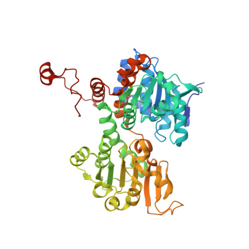
    We report biochemical and structural studies on inhibiting bacterial S -adenosyl-L-homocysteine hydrolase by transition metal cations. Our results revealed diverse molecular mechanisms of enzyme inactivation. Depending on the cation, the mechanism is based on arresting the enzyme in its closed, inactive conformation, disulfide bond formation within the active site or oxidation of the intermediate form of a cofactor.


  • Organizational Affiliation
    • Institute of Bioorganic Chemistry, Polish Academy of Sciences, Noskowskiego 12/14, 61-704 Poznan, Poland. kbrzezinski@ibch.poznan.pl.

Macromolecule Content 

  • Total Structure Weight: 213.8 kDa 
  • Atom Count: 16,326 
  • Modeled Residue Count: 1,843 
  • Deposited Residue Count: 1,888 
  • Unique protein chains: 1

Macromolecules

Find similar proteins by:|  3D Structure
Entity ID: 1
MoleculeChains  Sequence LengthOrganismDetailsImage
Adenosylhomocysteinase
A, B, C, D
472Pseudomonas aeruginosa PAO1Mutation(s): 0 
Gene Names: ahcYsahHPA0432
EC: 3.3.1.1 (PDB Primary Data), 3.13.2.1 (UniProt)
UniProt
Find proteins for Q9I685 (Pseudomonas aeruginosa (strain ATCC 15692 / DSM 22644 / CIP 104116 / JCM 14847 / LMG 12228 / 1C / PRS 101 / PAO1))
Explore Q9I685 
Go to UniProtKB:  Q9I685
Entity Groups
Sequence Clusters30% Identity50% Identity70% Identity90% Identity95% Identity100% Identity
UniProt GroupQ9I685
Sequence Annotations
Expand
Reference Sequence

Small Molecules

Ligands 8 Unique
IDChains Name / Formula / InChI Key2D Diagram3D Interactions
NAD
(Subject of Investigation/LOI)

Query on NAD


Download:Ideal Coordinates CCD File
E [auth A],
JA [auth D],
Q [auth B],
Z [auth C]
NICOTINAMIDE-ADENINE-DINUCLEOTIDE
C21 H27 N7 O14 P2
BAWFJGJZGIEFAR-NNYOXOHSSA-N
ADN
(Subject of Investigation/LOI)

Query on ADN


Download:Ideal Coordinates CCD File
AA [auth C],
F [auth A],
KA [auth D],
R [auth B]
ADENOSINE
C10 H13 N5 O4
OIRDTQYFTABQOQ-KQYNXXCUSA-N
HEZ

Query on HEZ


Download:Ideal Coordinates CCD File
S [auth B]HEXANE-1,6-DIOL
C6 H14 O2
XXMIOPMDWAUFGU-UHFFFAOYSA-N
CD
(Subject of Investigation/LOI)

Query on CD


Download:Ideal Coordinates CCD File
FA [auth C]
GA [auth C]
HA [auth C]
IA [auth C]
L [auth A]
FA [auth C],
GA [auth C],
HA [auth C],
IA [auth C],
L [auth A],
M [auth A],
N [auth A],
NA [auth D],
O [auth A],
OA [auth D],
P [auth A],
PA [auth D],
QA [auth D],
RA [auth D],
U [auth B],
V [auth B],
W [auth B],
X [auth B],
Y [auth B]
CADMIUM ION
Cd
WLZRMCYVCSSEQC-UHFFFAOYSA-N
BU1

Query on BU1


Download:Ideal Coordinates CCD File
BA [auth C]
CA [auth C]
DA [auth C]
G [auth A]
H [auth A]
BA [auth C],
CA [auth C],
DA [auth C],
G [auth A],
H [auth A],
LA [auth D]
1,4-BUTANEDIOL
C4 H10 O2
WERYXYBDKMZEQL-UHFFFAOYSA-N
PDO

Query on PDO


Download:Ideal Coordinates CCD File
I [auth A],
J [auth A]
1,3-PROPANDIOL
C3 H8 O2
YPFDHNVEDLHUCE-UHFFFAOYSA-N
K
(Subject of Investigation/LOI)

Query on K


Download:Ideal Coordinates CCD File
EA [auth C],
K [auth A],
MA [auth D],
T [auth B]
POTASSIUM ION
K
NPYPAHLBTDXSSS-UHFFFAOYSA-N
CL

Query on CL


Download:Ideal Coordinates CCD File
SA [auth D]CHLORIDE ION
Cl
VEXZGXHMUGYJMC-UHFFFAOYSA-M

Experimental Data & Validation

Experimental Data

  • Method: X-RAY DIFFRACTION
  • Resolution: 1.87 Å
  • R-Value Free:  0.162 (Depositor), 0.164 (DCC) 
  • R-Value Work:  0.147 (Depositor), 0.147 (DCC) 
  • R-Value Observed: 0.147 (Depositor) 
Space Group: C 1 2 1
Unit Cell:
Length ( Å )Angle ( ˚ )
a = 176.97α = 90
b = 134.16β = 106.063
c = 108.88γ = 90
Software Package:
Software NamePurpose
XDSdata reduction
XSCALEdata scaling
PHENIXrefinement
PDB_EXTRACTdata extraction
REFMACphasing

Structure Validation

View Full Validation Report


Ligand Structure Quality Assessment 


Entry History 

& Funding Information

Deposition Data


Funding OrganizationLocationGrant Number
Polish National Science CentrePolandSONATA BIS 2018/30/E/NZ1/00729

Revision History  (Full details and data files)

  • Version 1.0: 2023-04-19
    Type: Initial release
  • Version 1.1: 2024-02-07
    Changes: Data collection, Refinement description
  • Version 1.2: 2024-10-30
    Changes: Database references, Structure summary