7ZCW

Cryo-EM structure of GMPCPP-microtubules in complex with VASH2-SVBP


Experimental Data Snapshot

  • Method: ELECTRON MICROSCOPY
  • Resolution: 3.60 Å
  • Aggregation State: HELICAL ARRAY 
  • Reconstruction Method: SINGLE PARTICLE 

wwPDB Validation   3D Report Full Report


This is version 1.1 of the entry. See complete history


Literature

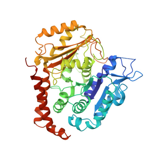
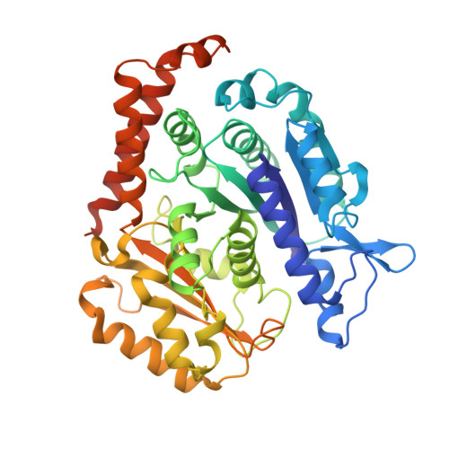
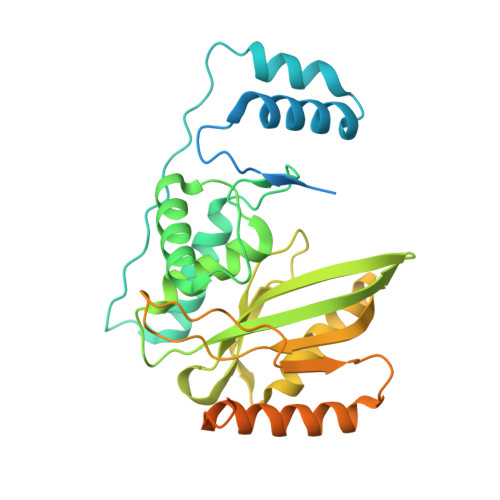
VASH1-SVBP and VASH2-SVBP generate different detyrosination profiles on microtubules.

Ramirez-Rios, S.Choi, S.R.Sanyal, C.Blum, T.B.Bosc, C.Krichen, F.Denarier, E.Soleilhac, J.M.Blot, B.Janke, C.Stoppin-Mellet, V.Magiera, M.M.Arnal, I.Steinmetz, M.O.Moutin, M.J.

(2023) J Cell Biol 222

  • DOI: https://doi.org/10.1083/jcb.202205096
  • Primary Citation of Related Structures:  
    7ZCW

  • PubMed Abstract: 

    The detyrosination/tyrosination cycle of α-tubulin is critical for proper cell functioning. VASH1-SVBP and VASH2-SVBP are ubiquitous enzymes involved in microtubule detyrosination, whose mode of action is little known. Here, we show in reconstituted systems and cells that VASH1-SVBP and VASH2-SVBP drive the global and local detyrosination of microtubules, respectively. We solved the cryo-electron microscopy structure of VASH2-SVBP bound to microtubules, revealing a different microtubule-binding configuration of its central catalytic region compared to VASH1-SVBP. We show that the divergent mode of detyrosination between the two enzymes is correlated with the microtubule-binding properties of their disordered N- and C-terminal regions. Specifically, the N-terminal region is responsible for a significantly longer residence time of VASH2-SVBP on microtubules compared to VASH1-SVBP. We suggest that this VASH region is critical for microtubule detachment and diffusion of VASH-SVBP enzymes on lattices. Our results suggest a mechanism by which VASH1-SVBP and VASH2-SVBP could generate distinct microtubule subpopulations and confined areas of detyrosinated lattices to drive various microtubule-based cellular functions.


  • Organizational Affiliation

    Univ. Grenoble Alpes, Inserm, U1216, Centre National de la Recherche Scientifique, Commissariat à l'Energie Atomique, Grenoble Institut Neurosciences, Grenoble, France.


Macromolecules
Find similar proteins by:  (by identity cutoff)  |  3D Structure
Entity ID: 1
MoleculeChains Sequence LengthOrganismDetailsImage
Tubulin alpha-1B chain
A, E
451Homo sapiensMutation(s): 0 
UniProt & NIH Common Fund Data Resources
Find proteins for P68363 (Homo sapiens)
Explore P68363 
Go to UniProtKB:  P68363
PHAROS:  P68363
GTEx:  ENSG00000123416 
Entity Groups  
Sequence Clusters30% Identity50% Identity70% Identity90% Identity95% Identity100% Identity
UniProt GroupP68363
Sequence Annotations
Expand
  • Reference Sequence
Find similar proteins by:  (by identity cutoff)  |  3D Structure
Entity ID: 2
MoleculeChains Sequence LengthOrganismDetailsImage
Tubulin beta-2B chain
B, F, G, H
445Homo sapiensMutation(s): 0 
UniProt & NIH Common Fund Data Resources
Find proteins for Q9BVA1 (Homo sapiens)
Explore Q9BVA1 
Go to UniProtKB:  Q9BVA1
PHAROS:  Q9BVA1
GTEx:  ENSG00000137285 
Entity Groups  
Sequence Clusters30% Identity50% Identity70% Identity90% Identity95% Identity100% Identity
UniProt GroupQ9BVA1
Sequence Annotations
Expand
  • Reference Sequence
Find similar proteins by:  (by identity cutoff)  |  3D Structure
Entity ID: 3
MoleculeChains Sequence LengthOrganismDetailsImage
Tubulinyl-Tyr carboxypeptidase 2355Homo sapiensMutation(s): 1 
Gene Names: VASH2VASHL
EC: 3.4.17.17
UniProt & NIH Common Fund Data Resources
Find proteins for Q86V25 (Homo sapiens)
Explore Q86V25 
Go to UniProtKB:  Q86V25
PHAROS:  Q86V25
GTEx:  ENSG00000143494 
Entity Groups  
Sequence Clusters30% Identity50% Identity70% Identity90% Identity95% Identity100% Identity
UniProt GroupQ86V25
Sequence Annotations
Expand
  • Reference Sequence
Find similar proteins by:  (by identity cutoff)  |  3D Structure
Entity ID: 4
MoleculeChains Sequence LengthOrganismDetailsImage
Small vasohibin-binding protein73Homo sapiensMutation(s): 0 
Gene Names: SVBPCCDC23
UniProt & NIH Common Fund Data Resources
Find proteins for Q8N300 (Homo sapiens)
Explore Q8N300 
Go to UniProtKB:  Q8N300
PHAROS:  Q8N300
GTEx:  ENSG00000177868 
Entity Groups  
Sequence Clusters30% Identity50% Identity70% Identity90% Identity95% Identity100% Identity
UniProt GroupQ8N300
Sequence Annotations
Expand
  • Reference Sequence
Small Molecules
Ligands 3 Unique
IDChains Name / Formula / InChI Key2D Diagram3D Interactions
GTP
Query on GTP

Download Ideal Coordinates CCD File 
I [auth A],
M [auth E]
GUANOSINE-5'-TRIPHOSPHATE
C10 H16 N5 O14 P3
XKMLYUALXHKNFT-UUOKFMHZSA-N
G2P
Query on G2P

Download Ideal Coordinates CCD File 
K [auth B],
O [auth F],
Q [auth G],
S [auth H]
PHOSPHOMETHYLPHOSPHONIC ACID GUANYLATE ESTER
C11 H18 N5 O13 P3
GXTIEXDFEKYVGY-KQYNXXCUSA-N
MG
Query on MG

Download Ideal Coordinates CCD File 
J [auth A]
L [auth B]
N [auth E]
P [auth F]
R [auth G]
J [auth A],
L [auth B],
N [auth E],
P [auth F],
R [auth G],
T [auth H]
MAGNESIUM ION
Mg
JLVVSXFLKOJNIY-UHFFFAOYSA-N
Experimental Data & Validation

Experimental Data

  • Method: ELECTRON MICROSCOPY
  • Resolution: 3.60 Å
  • Aggregation State: HELICAL ARRAY 
  • Reconstruction Method: SINGLE PARTICLE 
EM Software:
TaskSoftware PackageVersion
MODEL REFINEMENTPHENIX1.20.1-4487
MODEL REFINEMENTCoot0.9.7
RECONSTRUCTIONRELION3.0.8

Structure Validation

View Full Validation Report


Entry History & Funding Information

Deposition Data


Funding OrganizationLocationGrant Number
Swiss National Science FoundationSwitzerland310030_192566

Revision History  (Full details and data files)

  • Version 1.0: 2022-12-14
    Type: Initial release
  • Version 1.1: 2023-06-28
    Changes: Database references