7ZCN

Nitrite-bound MSOX movie series dataset 1 (0.8 MGy) of the copper nitrite reductase from Bradyrhizobium sp. ORS 375 (two-domain) - nitrite (start)


Experimental Data Snapshot

  • Method: X-RAY DIFFRACTION
  • Resolution: 1.19 Å
  • R-Value Free: 0.134 
  • R-Value Work: 0.108 
  • R-Value Observed: 0.109 

wwPDB Validation   3D Report Full Report


This is version 1.1 of the entry. See complete history


Literature

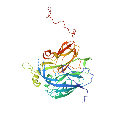
Single crystal spectroscopy and multiple structures from one crystal (MSOX) define catalysis in copper nitrite reductases.

Rose, S.L.Baba, S.Okumura, H.Antonyuk, S.V.Sasaki, D.Hedison, T.M.Shanmugam, M.Heyes, D.J.Scrutton, N.S.Kumasaka, T.Tosha, T.Eady, R.R.Yamamoto, M.Hasnain, S.S.

(2022) Proc Natl Acad Sci U S A 119: e2205664119-e2205664119

  • DOI: https://doi.org/10.1073/pnas.2205664119
  • Primary Citation of Related Structures:  
    7QXK, 7QY4, 7QYC, 7ZCN, 7ZCO, 7ZCP, 7ZCQ, 7ZCR, 7ZCS

  • PubMed Abstract: 

    Many enzymes utilize redox-coupled centers for performing catalysis where these centers are used to control and regulate the transfer of electrons required for catalysis, whose untimely delivery can lead to a state incapable of binding the substrate, i.e., a dead-end enzyme. Copper nitrite reductases (CuNiRs), which catalyze the reduction of nitrite to nitric oxide (NO), have proven to be a good model system for studying these complex processes including proton-coupled electron transfer (ET) and their orchestration for substrate binding/utilization. Recently, a two-domain CuNiR from a Rhizobia species ( Br 2D NiR) has been discovered with a substantially lower enzymatic activity where the catalytic type-2 Cu (T2Cu) site is occupied by two water molecules requiring their displacement for the substrate nitrite to bind. Single crystal spectroscopy combined with MSOX (multiple structures from one crystal) for both the as-isolated and nitrite-soaked crystals clearly demonstrate that inter-Cu ET within the coupled T1Cu-T2Cu redox system is heavily gated. Laser-flash photolysis and optical spectroscopy showed rapid ET from photoexcited NADH to the T1Cu center but little or no inter-Cu ET in the absence of nitrite. Furthermore, incomplete reoxidation of the T1Cu site (∼20% electrons transferred) was observed in the presence of nitrite, consistent with a slow formation of NO species in the serial structures of the MSOX movie obtained from the nitrite-soaked crystal, which is likely to be responsible for the lower activity of this CuNiR. Our approach is of direct relevance for studying redox reactions in a wide range of biological systems including metalloproteins that make up at least 30% of all proteins.


  • Organizational Affiliation

    Molecular Biophysics Group, Life Sciences Building, Institute of Systems, Molecular and Integrative Biology, Faculty of Health and Life Sciences, University of Liverpool, Liverpool, L69 7ZB, United Kingdom.


Macromolecules
Find similar proteins by:  (by identity cutoff)  |  3D Structure
Entity ID: 1
MoleculeChains Sequence LengthOrganismDetailsImage
Copper-containing nitrite reductase350Bradyrhizobium sp. ORS 375Mutation(s): 0 
Gene Names: nirKBRAO375_4030011
EC: 1.7.2.1
UniProt
Find proteins for H0SLX7 (Bradyrhizobium sp. (strain ORS 375))
Explore H0SLX7 
Go to UniProtKB:  H0SLX7
Entity Groups  
Sequence Clusters30% Identity50% Identity70% Identity90% Identity95% Identity100% Identity
UniProt GroupH0SLX7
Sequence Annotations
Expand
  • Reference Sequence
Oligosaccharides

Help

Entity ID: 2
MoleculeChains Length2D Diagram Glycosylation3D Interactions
beta-D-fructofuranose-(2-1)-alpha-D-glucopyranoseB [auth E],
C [auth F]
2N/A
Glycosylation Resources
GlyTouCan:  G05551OP
GlyCosmos:  G05551OP
Small Molecules
Ligands 4 Unique
IDChains Name / Formula / InChI Key2D Diagram3D Interactions
SO4
Query on SO4

Download Ideal Coordinates CCD File 
D [auth A]
E [auth A]
F [auth A]
G [auth A]
N [auth A]
D [auth A],
E [auth A],
F [auth A],
G [auth A],
N [auth A],
O [auth A],
P [auth A],
Q [auth A],
R [auth A],
S [auth A],
T [auth A]
SULFATE ION
O4 S
QAOWNCQODCNURD-UHFFFAOYSA-L
GOL
Query on GOL

Download Ideal Coordinates CCD File 
K [auth A],
L [auth A],
M [auth A]
GLYCEROL
C3 H8 O3
PEDCQBHIVMGVHV-UHFFFAOYSA-N
CU (Subject of Investigation/LOI)
Query on CU

Download Ideal Coordinates CCD File 
H [auth A],
I [auth A]
COPPER (II) ION
Cu
JPVYNHNXODAKFH-UHFFFAOYSA-N
NO2 (Subject of Investigation/LOI)
Query on NO2

Download Ideal Coordinates CCD File 
J [auth A],
U [auth A],
V [auth A],
W [auth A]
NITRITE ION
N O2
IOVCWXUNBOPUCH-UHFFFAOYSA-M
Biologically Interesting Molecules (External Reference) 1 Unique
Experimental Data & Validation

Experimental Data

  • Method: X-RAY DIFFRACTION
  • Resolution: 1.19 Å
  • R-Value Free: 0.134 
  • R-Value Work: 0.108 
  • R-Value Observed: 0.109 
  • Space Group: P 21 3
Unit Cell:
Length ( Å )Angle ( ˚ )
a = 106.725α = 90
b = 106.725β = 90
c = 106.725γ = 90
Software Package:
Software NamePurpose
REFMACrefinement
xia2data reduction
xia2data scaling
REFMACphasing

Structure Validation

View Full Validation Report


Entry History & Funding Information

Deposition Data


Funding OrganizationLocationGrant Number
UK Research and Innovation (UKRI)United KingdomBB/N013972/1
UK Research and Innovation (UKRI)United KingdomBB/S020055/1

Revision History  (Full details and data files)

  • Version 1.0: 2022-08-03
    Type: Initial release
  • Version 1.1: 2024-01-31
    Changes: Data collection, Refinement description