


yxBC from Bacillus subtilis in complex with Mn and N-oxalylglycine (NOG)
Chowdhury, R., Schofield, C.J.To be published.
Experimental Data Snapshot
Starting Model: experimental
View more details
Entity ID: 1 | |||||
|---|---|---|---|---|---|
| Molecule | Chains | Sequence Length | Organism | Details | Image |
| Uncharacterized protein YxbC | 341 | Bacillus subtilis subsp. subtilis str. 168 | Mutation(s): 0 Gene Names: yxbC, yxaQ, BSU39880, VE7D | 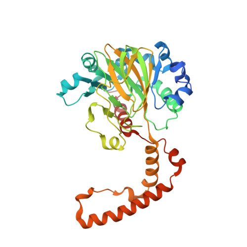 | |
UniProt | |||||
Entity Groups | |||||
| Sequence Clusters | 30% Identity50% Identity70% Identity90% Identity95% Identity100% Identity | ||||
| UniProt Group | P46327 | ||||
Sequence AnnotationsExpand | |||||
Reference Sequence | |||||
| Ligands 4 Unique | |||||
|---|---|---|---|---|---|
| ID | Chains | Name / Formula / InChI Key | 2D Diagram | 3D Interactions | |
| OGA (Subject of Investigation/LOI) Download:Ideal Coordinates CCD File | CA [auth D], F [auth A], O [auth B], U [auth C] | N-OXALYLGLYCINE C4 H5 N O5 BIMZLRFONYSTPT-UHFFFAOYSA-N |  | ||
| SO4 Download:Ideal Coordinates CCD File | AA [auth C] IA [auth D] JA [auth D] KA [auth D] L [auth A] | SULFATE ION O4 S QAOWNCQODCNURD-UHFFFAOYSA-L |  | ||
| GOL Download:Ideal Coordinates CCD File | DA [auth D] EA [auth D] FA [auth D] G [auth A] GA [auth D] | GLYCEROL C3 H8 O3 PEDCQBHIVMGVHV-UHFFFAOYSA-N |  | ||
| MN Download:Ideal Coordinates CCD File | BA [auth D], E [auth A], N [auth B], T [auth C] | MANGANESE (II) ION Mn WAEMQWOKJMHJLA-UHFFFAOYSA-N |  | ||
| Length ( Å ) | Angle ( ˚ ) |
|---|---|
| a = 109.733 | α = 90 |
| b = 109.733 | β = 90 |
| c = 192.191 | γ = 120 |
| Software Name | Purpose |
|---|---|
| SCALEPACK | data scaling |
| PHENIX | refinement |
| PDB_EXTRACT | data extraction |
| HKL-2000 | data reduction |
| PHASER | phasing |
| Funding Organization | Location | Grant Number |
|---|---|---|
| Not funded | -- |


