7ZAM | pdb_00007zam

Sam68


Experimental Data Snapshot

  • Method: X-RAY DIFFRACTION
  • Resolution: 2.79 Å
  • R-Value Free: 
    0.214 (Depositor), 0.197 (DCC) 
  • R-Value Work: 
    0.186 (Depositor) 

Starting Model: experimental
View more details

wwPDB Validation 3D Report Full Report

Validation slider image for 7ZAM

This is version 1.4 of the entry. See complete history

Literature

Structure and function analysis of Sam68 and hnRNP A1 synergy in the exclusion of exon 7 from SMN2 transcripts.

Nadal, M.Anton, R.Dorca-Arevalo, J.Estebanez-Perpina, E.Tizzano, E.F.Fuentes-Prior, P.

(2023) Protein Sci 32: e4553-e4553

  • DOI: https://doi.org/10.1002/pro.4553
  • Primary Citation Related Structures: 
    7Z89, 7Z8A, 7Z9A, 7Z9B, 7ZAB, 7ZAC, 7ZAF, 7ZAM

  • PubMed Abstract: 

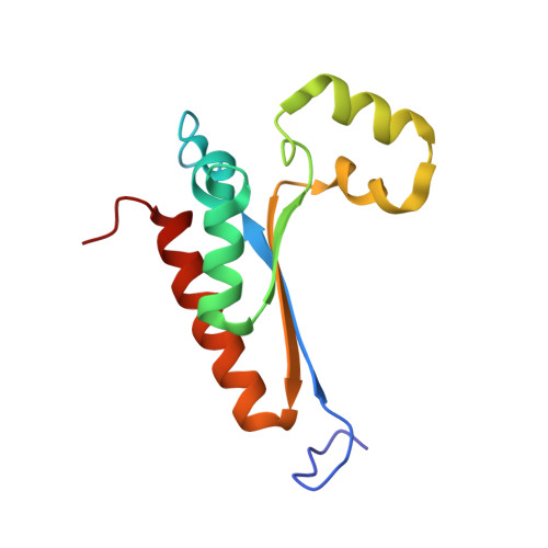
    Spinal muscular atrophy (SMA) is a neurodegenerative disease caused by the absence of a functional copy of the Survival of Motor Neuron 1 gene (SMN1). The nearly identical paralog, SMN2, cannot compensate for the loss of SMN1 because exon 7 is aberrantly skipped from most SMN2 transcripts, a process mediated by synergistic activities of Src-associated during mitosis, 68 kDa (Sam68/KHDRBS1) and heterogeneous nuclear ribonucleoprotein (hnRNP) A1. This results in the production of a truncated, nonfunctional protein that is rapidly degraded. Here, we present several crystal structures of Sam68 RNA-binding domain (RBD). Sam68-RBD forms stable symmetric homodimers by antiparallel association of helices α3 from two monomers. However, the details of domain organization and the dimerization interface differ significantly from previously characterized homologs. We demonstrate that Sam68 and hnRNP A1 can simultaneously bind proximal motifs within the central region of SMN2 (ex7). Furthermore, we show that the RNA-binding pockets of the two proteins are close to each other in their heterodimeric complex and identify contact residues using crosslinking-mass spectrometry. We present a model of the ternary Sam68·SMN2 (ex7)·hnRNP A1 complex that reconciles all available information on SMN1/2 splicing. Our findings have important implications for the etiology of SMA and open new avenues for the design of novel therapeutics to treat splicing diseases.


  • Organizational Affiliation
    • Molecular Bases of Disease, Biomedical Research Institute Sant Pau (IIB Sant Pau), Barcelona, Spain.

Macromolecule Content 

  • Total Structure Weight: 28.45 kDa 
  • Atom Count: 1,991 
  • Modeled Residue Count: 236 
  • Deposited Residue Count: 236 
  • Unique protein chains: 1

Macromolecules

Find similar proteins by:|  3D Structure
Entity ID: 1
MoleculeChains  Sequence LengthOrganismDetailsImage
Isoform 2 of KH domain-containing, RNA-binding, signal transduction-associated protein 1A [auth AAA],
B [auth BBB]
118Homo sapiensMutation(s): 1 
Gene Names: KHDRBS1SAM68
UniProt & NIH Common Fund Data Resources
Find proteins for Q07666 (Homo sapiens)
Explore Q07666 
Go to UniProtKB:  Q07666
PHAROS:  Q07666
GTEx:  ENSG00000121774 
Entity Groups
Sequence Clusters30% Identity50% Identity70% Identity90% Identity95% Identity100% Identity
UniProt GroupQ07666
Sequence Annotations
Expand
Reference Sequence

Small Molecules

Ligands 4 Unique
IDChains Name / Formula / InChI Key2D Diagram3D Interactions
IOD

Query on IOD


Download:Ideal Coordinates CCD File
C [auth AAA],
O [auth BBB]
IODIDE ION
I
XMBWDFGMSWQBCA-UHFFFAOYSA-M
SO4

Query on SO4


Download:Ideal Coordinates CCD File
P [auth BBB],
Q [auth BBB]
SULFATE ION
O4 S
QAOWNCQODCNURD-UHFFFAOYSA-L
GOL

Query on GOL


Download:Ideal Coordinates CCD File
N [auth AAA]GLYCEROL
C3 H8 O3
PEDCQBHIVMGVHV-UHFFFAOYSA-N
EDO

Query on EDO


Download:Ideal Coordinates CCD File
D [auth AAA]
E [auth AAA]
F [auth AAA]
G [auth AAA]
H [auth AAA]
D [auth AAA],
E [auth AAA],
F [auth AAA],
G [auth AAA],
H [auth AAA],
I [auth AAA],
J [auth AAA],
K [auth AAA],
L [auth AAA],
M [auth AAA],
R [auth BBB],
S [auth BBB],
T [auth BBB],
U [auth BBB],
V [auth BBB]
1,2-ETHANEDIOL
C2 H6 O2
LYCAIKOWRPUZTN-UHFFFAOYSA-N

Experimental Data & Validation

Experimental Data

  • Method: X-RAY DIFFRACTION
  • Resolution: 2.79 Å
  • R-Value Free:  0.214 (Depositor), 0.197 (DCC) 
  • R-Value Work:  0.186 (Depositor) 
Space Group: C 2 2 21
Unit Cell:
Length ( Å )Angle ( ˚ )
a = 138.459α = 90
b = 138.701β = 90
c = 82.337γ = 90
Software Package:
Software NamePurpose
REFMACrefinement
XDSdata reduction
Aimlessdata scaling
MOLREPphasing

Structure Validation

View Full Validation Report


Entry History 

& Funding Information

Deposition Data


Funding OrganizationLocationGrant Number
Other private--

Revision History  (Full details and data files)

  • Version 1.0: 2023-02-01
    Type: Initial release
  • Version 1.1: 2023-02-08
    Changes: Derived calculations
  • Version 1.2: 2023-04-12
    Changes: Database references
  • Version 1.3: 2024-02-07
    Changes: Data collection, Refinement description
  • Version 1.4: 2024-11-13
    Changes: Structure summary