7Z88

DNA-PK in the intermediate state


Experimental Data Snapshot

  • Method: ELECTRON MICROSCOPY
  • Resolution: 3.33 Å
  • Aggregation State: PARTICLE 
  • Reconstruction Method: SINGLE PARTICLE 

wwPDB Validation   3D Report Full Report


This is version 1.1 of the entry. See complete history


Literature

Human DNA-dependent protein kinase activation mechanism.

Liang, S.Blundell, T.L.

(2023) Nat Struct Mol Biol 30: 140-147

  • DOI: https://doi.org/10.1038/s41594-022-00881-w
  • Primary Citation of Related Structures:  
    7Z87, 7Z88

  • PubMed Abstract: 

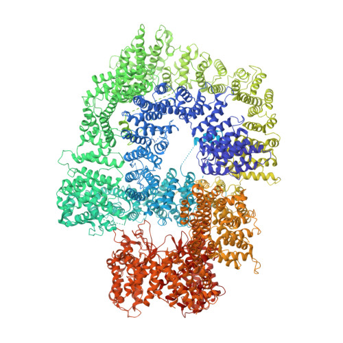
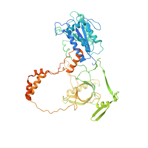
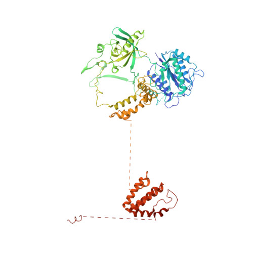
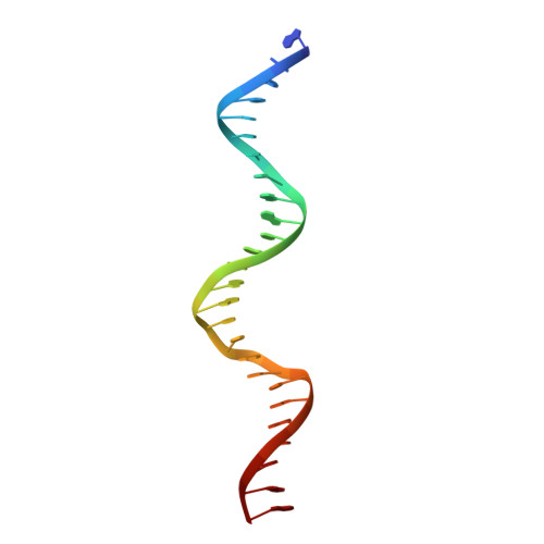
    DNA-dependent protein kinase (DNA-PK), a multicomponent complex including the DNA-PK catalytic subunit and Ku70/80 heterodimer together with DNA, is central to human DNA damage response and repair. Using a DNA-PK-selective inhibitor (M3814), we identified from one dataset two cryo-EM structures of the human DNA-PK complex in different states, the intermediate state and the active state. Here we show that activation of the kinase is regulated through conformational changes caused by the binding ligand and the string region (residues 802-846) of the DNA-PK catalytic subunit, particularly the helix-hairpin-helix motif (residues 816-836) that interacts with DNA. These observations demonstrate the regulatory role of the ligand and explain why DNA-PK is DNA dependent. Cooperation and coordination among binding partners, disordered flexible regions and mechanically flexible HEAT repeats modulate the activation of the kinase. Together with previous findings, these results provide a better molecular understanding of DNA-PK catalysis.


  • Organizational Affiliation

    Department of Biochemistry, University of Cambridge, Cambridge, UK. sl744@cam.ac.uk.


Macromolecules

Find similar proteins by:  (by identity cutoff)  |  3D Structure
Entity ID: 1
MoleculeChains Sequence LengthOrganismDetailsImage
DNA-dependent protein kinase catalytic subunit4,128Homo sapiensMutation(s): 0 
EC: 2.7.11.1
UniProt & NIH Common Fund Data Resources
Find proteins for P78527 (Homo sapiens)
Explore P78527 
Go to UniProtKB:  P78527
PHAROS:  P78527
GTEx:  ENSG00000253729 
Entity Groups  
Sequence Clusters30% Identity50% Identity70% Identity90% Identity95% Identity100% Identity
UniProt GroupP78527
Sequence Annotations
Expand
  • Reference Sequence
Find similar proteins by:  (by identity cutoff)  |  3D Structure
Entity ID: 2
MoleculeChains Sequence LengthOrganismDetailsImage
X-ray repair cross-complementing protein 6609Homo sapiensMutation(s): 0 
Gene Names: XRCC6G22P1
EC: 3.6.4 (PDB Primary Data), 4.2.99 (PDB Primary Data)
UniProt & NIH Common Fund Data Resources
Find proteins for P12956 (Homo sapiens)
Explore P12956 
Go to UniProtKB:  P12956
PHAROS:  P12956
GTEx:  ENSG00000196419 
Entity Groups  
Sequence Clusters30% Identity50% Identity70% Identity90% Identity95% Identity100% Identity
UniProt GroupP12956
Sequence Annotations
Expand
  • Reference Sequence
Find similar proteins by:  (by identity cutoff)  |  3D Structure
Entity ID: 3
MoleculeChains Sequence LengthOrganismDetailsImage
X-ray repair cross-complementing protein 5732Homo sapiensMutation(s): 0 
Gene Names: XRCC5G22P2
EC: 3.6.4
UniProt & NIH Common Fund Data Resources
Find proteins for P13010 (Homo sapiens)
Explore P13010 
Go to UniProtKB:  P13010
PHAROS:  P13010
GTEx:  ENSG00000079246 
Entity Groups  
Sequence Clusters30% Identity50% Identity70% Identity90% Identity95% Identity100% Identity
UniProt GroupP13010
Sequence Annotations
Expand
  • Reference Sequence
Find similar nucleic acids by:  (by identity cutoff)  |  3D Structure
Entity ID: 4
MoleculeChains LengthOrganismImage
DNA (26-MER)26Homo sapiens
Sequence Annotations
Expand
  • Reference Sequence
Find similar nucleic acids by:  (by identity cutoff)  |  3D Structure
Entity ID: 5
MoleculeChains LengthOrganismImage
DNA (26-MER)26Homo sapiens
Sequence Annotations
Expand
  • Reference Sequence
Small Molecules
Ligands 1 Unique
IDChains Name / Formula / InChI Key2D Diagram3D Interactions
1IX (Subject of Investigation/LOI)
Query on 1IX

Download Ideal Coordinates CCD File 
F [auth A](~{S})-[2-chloranyl-4-fluoranyl-5-(7-morpholin-4-ylquinazolin-4-yl)phenyl]-(6-methoxypyridazin-3-yl)methanol
C24 H21 Cl F N5 O3
MOWXJLUYGFNTAL-DEOSSOPVSA-N
Experimental Data & Validation

Experimental Data

  • Method: ELECTRON MICROSCOPY
  • Resolution: 3.33 Å
  • Aggregation State: PARTICLE 
  • Reconstruction Method: SINGLE PARTICLE 

Structure Validation

View Full Validation Report


Entry History & Funding Information

Deposition Data


Funding OrganizationLocationGrant Number
Wellcome TrustUnited Kingdom--

Revision History  (Full details and data files)

  • Version 1.0: 2023-01-18
    Type: Initial release
  • Version 1.1: 2023-03-01
    Changes: Database references