7Z5S

Crystal Structure of botulinum neurotoxin A2 cell binding domain in complex with GD1a


Experimental Data Snapshot

  • Method: X-RAY DIFFRACTION
  • Resolution: 2.10 Å
  • R-Value Free: 0.226 
  • R-Value Work: 0.188 

wwPDB Validation   3D Report Full Report


This is version 1.1 of the entry. See complete history


Literature

Structural Features of Clostridium botulinum Neurotoxin Subtype A2 Cell Binding Domain.

Gregory, K.S.Mahadeva, T.B.Liu, S.M.Acharya, K.R.

(2022) Toxins (Basel) 14

  • DOI: https://doi.org/10.3390/toxins14050356
  • Primary Citation of Related Structures:  
    7Z5S, 7Z5T

  • PubMed Abstract: 

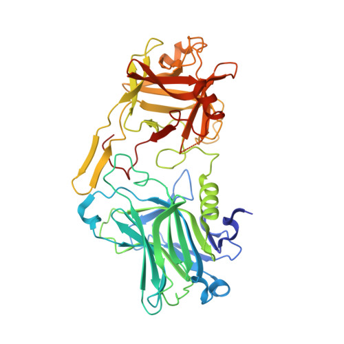
    Botulinum neurotoxins (BoNT) are a group of clostridial toxins that cause the potentially fatal neuroparalytic disease botulism. Although highly toxic, BoNTs are utilized as therapeutics to treat a range of neuromuscular conditions. Several serotypes (BoNT/A-/G, /X) have been identified with vastly differing toxicological profiles. Each serotype can be further sub-categorised into subtypes due to subtle variations in their protein sequence. These minor changes have been attributed to differences in both the duration of action and potency for BoNT/A subtypes. BoNTs are composed of three domains-a cell-binding domain, a translocation domain, and a catalytic domain. In this paper, we present the crystal structures of the botulinum neurotoxin A2 cell binding domain, both alone and in complex with its receptor ganglioside GD1a at 1.63 and 2.10 Å, respectively. The analysis of these structures reveals a potential redox-dependent Lys-O-Cys bridge close to the ganglioside binding site and a hinge motion between the H CN and H CC subdomains. Furthermore, we make a detailed comparison with the previously reported H C /A2:SV2C structure for a comprehensive structural analysis of H C /A2 receptor binding.


  • Organizational Affiliation

    Department of Biology and Biochemistry, University of Bath, Claverton Down, Bath BA2 7AY, UK.


Macromolecules
Find similar proteins by:  (by identity cutoff)  |  3D Structure
Entity ID: 1
MoleculeChains Sequence LengthOrganismDetailsImage
Botulinum neurotoxinA [auth AAA],
B [auth BBB]
433Clostridium botulinumMutation(s): 0 
Gene Names: Abont
UniProt
Find proteins for K4GGE0 (Clostridium botulinum)
Explore K4GGE0 
Go to UniProtKB:  K4GGE0
Entity Groups  
Sequence Clusters30% Identity50% Identity70% Identity90% Identity95% Identity100% Identity
UniProt GroupK4GGE0
Sequence Annotations
Expand
  • Reference Sequence
Oligosaccharides

Help

Entity ID: 2
MoleculeChains Length2D Diagram Glycosylation3D Interactions
N-acetyl-alpha-neuraminic acid-(2-3)-beta-D-galactopyranose-(1-3)-2-acetamido-2-deoxy-beta-D-galactopyranose-(1-4)-[N-acetyl-alpha-neuraminic acid-(2-3)]beta-D-galactopyranoseC [auth DDD]5N/A
Glycosylation Resources
GlyTouCan:  G78641QJ
GlyCosmos:  G78641QJ
GlyGen:  G78641QJ
Experimental Data & Validation

Experimental Data

  • Method: X-RAY DIFFRACTION
  • Resolution: 2.10 Å
  • R-Value Free: 0.226 
  • R-Value Work: 0.188 
  • Space Group: P 41
Unit Cell:
Length ( Å )Angle ( ˚ )
a = 105.068α = 90
b = 105.068β = 90
c = 132.58γ = 90
Software Package:
Software NamePurpose
REFMACrefinement
DIALSdata reduction
Aimlessdata scaling
PHASERphasing

Structure Validation

View Full Validation Report


Entry History & Funding Information

Deposition Data


Funding OrganizationLocationGrant Number
Not funded--

Revision History  (Full details and data files)

  • Version 1.0: 2022-06-08
    Type: Initial release
  • Version 1.1: 2024-01-31
    Changes: Data collection, Derived calculations, Refinement description