7Z5O

W-formate dehydrogenase from Desulfovibrio vulgaris - Dithionite reduced form


Experimental Data Snapshot

  • Method: X-RAY DIFFRACTION
  • Resolution: 1.53 Å
  • R-Value Free: 0.153 
  • R-Value Work: 0.119 

wwPDB Validation   3D Report Full Report


Ligand Structure Quality Assessment 


This is version 2.2 of the entry. See complete history


Literature

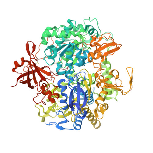
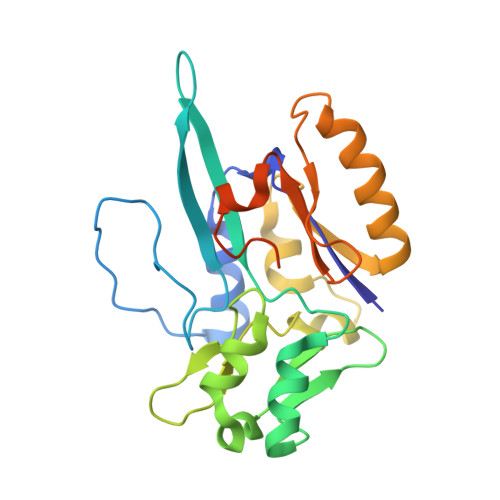
Spectroscopic and Structural Characterization of Reduced Desulfovibrio vulgaris Hildenborough W-FdhAB Reveals Stable Metal Coordination during Catalysis.

Oliveira, A.R.Mota, C.Klymanska, K.Biaso, F.Romao, M.J.Guigliarelli, B.Pereira, I.C.

(2022) ACS Chem Biol 17: 1901-1909

  • DOI: https://doi.org/10.1021/acschembio.2c00336
  • Primary Citation of Related Structures:  
    7Z5O

  • PubMed Abstract: 

    Metal-dependent formate dehydrogenases are important enzymes due to their activity of CO 2 reduction to formate. The tungsten-containing FdhAB formate dehydrogenase from Desulfovibrio vulgaris Hildenborough is a good example displaying high activity, simple composition, and a notable structural and catalytic robustness. Here, we report the first spectroscopic redox characterization of FdhAB metal centers by EPR. Titration with dithionite or formate leads to reduction of three [4Fe-4S] 1+ clusters, and full reduction requires Ti(III)-citrate. The redox potentials of the four [4Fe-4S] 1+ centers range between -250 and -530 mV. Two distinct W V signals were detected, W D V and W F V , which differ in only the g 2 -value. This difference can be explained by small variations in the twist angle of the two pyranopterins, as determined through DFT calculations of model compounds. The redox potential of W VI/V was determined to be -370 mV when reduced by dithionite and -340 mV when reduced by formate. The crystal structure of dithionite-reduced FdhAB was determined at high resolution (1.5 Å), revealing the same structural alterations as reported for the formate-reduced structure. These results corroborate a stable six-ligand W coordination in the catalytic intermediate W V state of FdhAB.


  • Organizational Affiliation

    Instituto de Tecnologia Química e Biológica António Xavier, Universidade Nova de Lisboa, Av. da República, 2780-157 Oeiras, Portugal.


Macromolecules
Find similar proteins by:  (by identity cutoff)  |  3D Structure
Entity ID: 1
MoleculeChains Sequence LengthOrganismDetailsImage
Formate dehydrogenase, alpha subunit, selenocysteine-containingA [auth AAA]1,009Nitratidesulfovibrio vulgaris str. HildenboroughMutation(s): 0 
Gene Names: fdnG-1DVU_0587
EC: 1.2.1.2
UniProt
Find proteins for Q72EJ1 (Desulfovibrio vulgaris (strain ATCC 29579 / DSM 644 / NCIMB 8303 / VKM B-1760 / Hildenborough))
Explore Q72EJ1 
Go to UniProtKB:  Q72EJ1
Entity Groups  
Sequence Clusters30% Identity50% Identity70% Identity90% Identity95% Identity100% Identity
UniProt GroupQ72EJ1
Sequence Annotations
Expand
  • Reference Sequence
Find similar proteins by:  (by identity cutoff)  |  3D Structure
Entity ID: 2
MoleculeChains Sequence LengthOrganismDetailsImage
Formate dehydrogenase, beta subunit, putativeB [auth BBB]236Nitratidesulfovibrio vulgaris str. HildenboroughMutation(s): 0 
Gene Names: DVU_0588
UniProt
Find proteins for Q72EJ0 (Desulfovibrio vulgaris (strain ATCC 29579 / DSM 644 / NCIMB 8303 / VKM B-1760 / Hildenborough))
Explore Q72EJ0 
Go to UniProtKB:  Q72EJ0
Entity Groups  
Sequence Clusters30% Identity50% Identity70% Identity90% Identity95% Identity100% Identity
UniProt GroupQ72EJ0
Sequence Annotations
Expand
  • Reference Sequence
Small Molecules
Ligands 7 Unique
IDChains Name / Formula / InChI Key2D Diagram3D Interactions
MGD (Subject of Investigation/LOI)
Query on MGD

Download Ideal Coordinates CCD File 
C [auth AAA],
D [auth AAA]
2-AMINO-5,6-DIMERCAPTO-7-METHYL-3,7,8A,9-TETRAHYDRO-8-OXA-1,3,9,10-TETRAAZA-ANTHRACEN-4-ONE GUANOSINE DINUCLEOTIDE
C20 H26 N10 O13 P2 S2
VQAGYJCYOLHZDH-ILXWUORBSA-N
SF4
Query on SF4

Download Ideal Coordinates CCD File 
E [auth AAA],
S [auth BBB],
T [auth BBB],
U [auth BBB]
IRON/SULFUR CLUSTER
Fe4 S4
LJBDFODJNLIPKO-UHFFFAOYSA-N
W (Subject of Investigation/LOI)
Query on W

Download Ideal Coordinates CCD File 
G [auth AAA]TUNGSTEN ION
W
FZFRVZDLZISPFJ-UHFFFAOYSA-N
PEG
Query on PEG

Download Ideal Coordinates CCD File 
Q [auth AAA]DI(HYDROXYETHYL)ETHER
C4 H10 O3
MTHSVFCYNBDYFN-UHFFFAOYSA-N
GOL
Query on GOL

Download Ideal Coordinates CCD File 
H [auth AAA]
I [auth AAA]
J [auth AAA]
K [auth AAA]
L [auth AAA]
H [auth AAA],
I [auth AAA],
J [auth AAA],
K [auth AAA],
L [auth AAA],
M [auth AAA],
N [auth AAA],
P [auth AAA]
GLYCEROL
C3 H8 O3
PEDCQBHIVMGVHV-UHFFFAOYSA-N
NO3
Query on NO3

Download Ideal Coordinates CCD File 
O [auth AAA],
R [auth AAA]
NITRATE ION
N O3
NHNBFGGVMKEFGY-UHFFFAOYSA-N
H2S (Subject of Investigation/LOI)
Query on H2S

Download Ideal Coordinates CCD File 
F [auth AAA]HYDROSULFURIC ACID
H2 S
RWSOTUBLDIXVET-UHFFFAOYSA-N
Experimental Data & Validation

Experimental Data

  • Method: X-RAY DIFFRACTION
  • Resolution: 1.53 Å
  • R-Value Free: 0.153 
  • R-Value Work: 0.119 
  • Space Group: P 21 21 21
Unit Cell:
Length ( Å )Angle ( ˚ )
a = 64.811α = 90
b = 124.15β = 90
c = 149.666γ = 90
Software Package:
Software NamePurpose
REFMACrefinement
autoPROCdata processing
PHASERphasing

Structure Validation

View Full Validation Report


Ligand Structure Quality Assessment 


Entry History & Funding Information

Deposition Data


Funding OrganizationLocationGrant Number
Fundacao para a Ciencia e a TecnologiaPortugalPTDC/BII-BBF/2050/2020

Revision History  (Full details and data files)

  • Version 1.0: 2022-07-13
    Type: Initial release
  • Version 2.0: 2022-07-20
    Type: Coordinate replacement
    Reason: Atomic clashes
    Changes: Advisory, Atomic model, Author supporting evidence, Data collection, Derived calculations, Refinement description
  • Version 2.1: 2022-07-27
    Changes: Database references
  • Version 2.2: 2024-01-31
    Changes: Data collection, Refinement description