7Z3R | pdb_00007z3r

Crystal structure of the mouse leptin:LepR-IgCRH2 complex to 2.95 A resolution.


Experimental Data Snapshot

  • Method: X-RAY DIFFRACTION
  • Resolution: 2.95 Å
  • R-Value Free: 
    0.251 (Depositor), 0.242 (DCC) 
  • R-Value Work: 
    0.223 (Depositor), 0.210 (DCC) 
  • R-Value Observed: 
    0.225 (Depositor) 

Starting Model: experimental
View more details

wwPDB Validation 3D Report Full Report

Validation slider image for 7Z3R

Ligand Structure Quality Assessment 


This is version 1.4 of the entry. See complete history

Literature

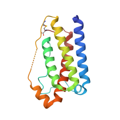
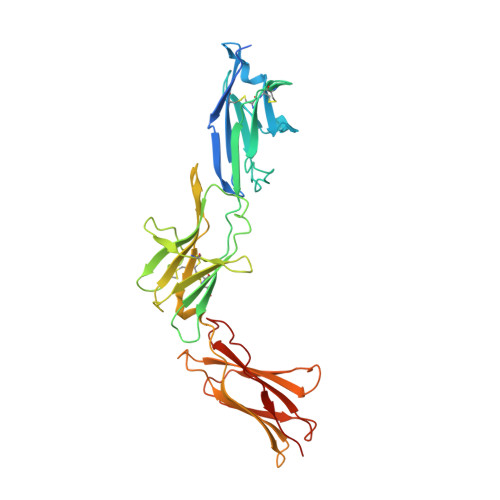
Mechanism of receptor assembly via the pleiotropic adipokine Leptin.

Tsirigotaki, A.Dansercoer, A.Verschueren, K.H.G.Markovic, I.Pollmann, C.Hafer, M.Felix, J.Birck, C.Van Putte, W.Catteeuw, D.Tavernier, J.Fernando Bazan, J.Piehler, J.Savvides, S.N.Verstraete, K.

(2023) Nat Struct Mol Biol 30: 551-563

  • DOI: https://doi.org/10.1038/s41594-023-00941-9
  • Primary Citation Related Structures: 
    7Z3P, 7Z3Q, 7Z3R, 8AV2, 8AVB, 8AVC, 8AVD, 8AVE, 8AVF, 8AVO, 8B7Q

  • PubMed Abstract: 

    The adipokine Leptin activates its receptor LEP-R in the hypothalamus to regulate body weight and exerts additional pleiotropic functions in immunity, fertility and cancer. However, the structure and mechanism of Leptin-mediated LEP-R assemblies has remained unclear. Intriguingly, the signaling-competent isoform of LEP-R is only lowly abundant amid several inactive short LEP-R isoforms contributing to a mechanistic conundrum. Here we show by X-ray crystallography and cryo-EM that, in contrast to long-standing paradigms, Leptin induces type I cytokine receptor assemblies featuring 3:3 stoichiometry and demonstrate such Leptin-induced trimerization of LEP-R on living cells via single-molecule microscopy. In mediating these assemblies, Leptin undergoes drastic restructuring that activates its site III for binding to the Ig domain of an adjacent LEP-R. These interactions are abolished by mutations linked to obesity. Collectively, our study provides the structural and mechanistic framework for how evolutionarily conserved Leptin:LEP-R assemblies with 3:3 stoichiometry can engage distinct LEP-R isoforms to achieve signaling.


  • Organizational Affiliation
    • Unit for Structural Biology, Department of Biochemistry and Microbiology, Ghent University, Ghent, Belgium.

Macromolecule Content 

  • Total Structure Weight: 53.87 kDa 
  • Atom Count: 3,563 
  • Modeled Residue Count: 447 
  • Deposited Residue Count: 478 
  • Unique protein chains: 2

Macromolecules

Find similar proteins by:|  3D Structure
Entity ID: 1
MoleculeChains  Sequence LengthOrganismDetailsImage
Leptin148Mus musculusMutation(s): 0 
Gene Names: LepOb
UniProt & NIH Common Fund Data Resources
Find proteins for P41160 (Mus musculus)
Explore P41160 
Go to UniProtKB:  P41160
IMPC:  MGI:104663
Entity Groups
Sequence Clusters30% Identity50% Identity70% Identity90% Identity95% Identity100% Identity
UniProt GroupP41160
Sequence Annotations
Expand
Reference Sequence
Find similar proteins by:|  3D Structure
Entity ID: 2
MoleculeChains  Sequence LengthOrganismDetailsImage
Leptin receptor330Mus musculusMutation(s): 1 
Gene Names: LeprDbObr
UniProt & NIH Common Fund Data Resources
Find proteins for P48356 (Mus musculus)
Explore P48356 
Go to UniProtKB:  P48356
IMPC:  MGI:104993
Entity Groups
Sequence Clusters30% Identity50% Identity70% Identity90% Identity95% Identity100% Identity
UniProt GroupP48356
Glycosylation
Glycosylation Sites: 1Go to GlyGen: P48356-1
Sequence Annotations
Expand
Reference Sequence

Small Molecules

Ligands 1 Unique
IDChains Name / Formula / InChI Key2D Diagram3D Interactions
NAG

Query on NAG


Download:Ideal Coordinates CCD File
C [auth B]2-acetamido-2-deoxy-beta-D-glucopyranose
C8 H15 N O6
OVRNDRQMDRJTHS-FMDGEEDCSA-N

Experimental Data & Validation

Experimental Data

  • Method: X-RAY DIFFRACTION
  • Resolution: 2.95 Å
  • R-Value Free:  0.251 (Depositor), 0.242 (DCC) 
  • R-Value Work:  0.223 (Depositor), 0.210 (DCC) 
  • R-Value Observed: 0.225 (Depositor) 
Space Group: H 3 2
Unit Cell:
Length ( Å )Angle ( ˚ )
a = 132.678α = 90
b = 132.678β = 90
c = 249.191γ = 120
Software Package:
Software NamePurpose
BUSTERrefinement
XDSdata reduction
Aimlessdata scaling
PHASERphasing

Structure Validation

View Full Validation Report


Ligand Structure Quality Assessment 


Entry History 

& Funding Information

Deposition Data


Funding OrganizationLocationGrant Number
Research Foundation - Flanders (FWO)BelgiumG0G0619N

Revision History  (Full details and data files)

  • Version 1.0: 2023-03-22
    Type: Initial release
  • Version 1.1: 2023-04-05
    Changes: Database references
  • Version 1.2: 2023-04-26
    Changes: Database references
  • Version 1.3: 2024-02-07
    Changes: Data collection, Refinement description
  • Version 1.4: 2024-11-06
    Changes: Structure summary