7Z3E

XFEL structure of Class Ib ribonucleotide reductase dimanganese(II) NrdF in complex with hydroquinone NrdI from Bacillus cereus


Experimental Data Snapshot

  • Method: X-RAY DIFFRACTION
  • Resolution: 2.00 Å
  • R-Value Free: 0.184 
  • R-Value Work: 0.152 
  • R-Value Observed: 0.153 

wwPDB Validation   3D Report Full Report


Ligand Structure Quality Assessment 


This is version 1.1 of the entry. See complete history


Literature

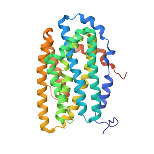
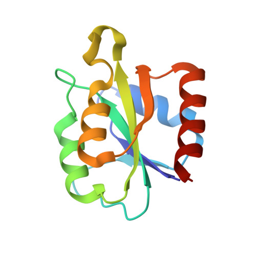
Redox-controlled reorganization and flavin strain within the ribonucleotide reductase R2b-NrdI complex monitored by serial femtosecond crystallography.

John, J.Aurelius, O.Srinivas, V.Saura, P.Kim, I.S.Bhowmick, A.Simon, P.S.Dasgupta, M.Pham, C.Gul, S.Sutherlin, K.D.Aller, P.Butryn, A.Orville, A.M.Cheah, M.H.Owada, S.Tono, K.Fuller, F.D.Batyuk, A.Brewster, A.S.Sauter, N.K.Yachandra, V.K.Yano, J.Kaila, V.R.I.Kern, J.Lebrette, H.Hogbom, M.

(2022) Elife 11

  • DOI: https://doi.org/10.7554/eLife.79226
  • Primary Citation of Related Structures:  
    7Z3D, 7Z3E

  • PubMed Abstract: 

    Redox reactions are central to biochemistry and are both controlled by and induce protein structural changes. Here, we describe structural rearrangements and crosstalk within the Bacillus cereus ribonucleotide reductase R2b-NrdI complex, a di-metal carboxylate-flavoprotein system, as part of the mechanism generating the essential catalytic free radical of the enzyme. Femtosecond crystallography at an X-ray free electron laser was utilized to obtain structures at room temperature in defined redox states without suffering photoreduction. Together with density functional theory calculations, we show that the flavin is under steric strain in the R2b-NrdI protein complex, likely tuning its redox properties to promote superoxide generation. Moreover, a binding site in close vicinity to the expected flavin O 2 interaction site is observed to be controlled by the redox state of the flavin and linked to the channel proposed to funnel the produced superoxide species from NrdI to the di-manganese site in protein R2b. These specific features are coupled to further structural changes around the R2b-NrdI interaction surface. The mechanistic implications for the control of reactive oxygen species and radical generation in protein R2b are discussed.


  • Organizational Affiliation

    Department of Biochemistry and Biophysics, Arrhenius Laboratories for Natural Sciences, Stockholm University, Stockholm, Sweden.


Macromolecules
Find similar proteins by:  (by identity cutoff)  |  3D Structure
Entity ID: 1
MoleculeChains Sequence LengthOrganismDetailsImage
Ribonucleoside-diphosphate reductase subunit beta322Bacillus cereus ATCC 14579Mutation(s): 0 
Gene Names: BC_1355
EC: 1.17.4.1
UniProt
Find proteins for Q81G55 (Bacillus cereus (strain ATCC 14579 / DSM 31 / CCUG 7414 / JCM 2152 / NBRC 15305 / NCIMB 9373 / NCTC 2599 / NRRL B-3711))
Explore Q81G55 
Go to UniProtKB:  Q81G55
Entity Groups  
Sequence Clusters30% Identity50% Identity70% Identity90% Identity95% Identity100% Identity
UniProt GroupQ81G55
Sequence Annotations
Expand
  • Reference Sequence
Find similar proteins by:  (by identity cutoff)  |  3D Structure
Entity ID: 2
MoleculeChains Sequence LengthOrganismDetailsImage
Protein NrdI119Bacillus cereus ATCC 14579Mutation(s): 0 
Gene Names: 
UniProt
Find proteins for Q81G57 (Bacillus cereus (strain ATCC 14579 / DSM 31 / CCUG 7414 / JCM 2152 / NBRC 15305 / NCIMB 9373 / NCTC 2599 / NRRL B-3711))
Explore Q81G57 
Go to UniProtKB:  Q81G57
Entity Groups  
Sequence Clusters30% Identity50% Identity70% Identity90% Identity95% Identity100% Identity
UniProt GroupQ81G57
Sequence Annotations
Expand
  • Reference Sequence
Experimental Data & Validation

Experimental Data

  • Method: X-RAY DIFFRACTION
  • Resolution: 2.00 Å
  • R-Value Free: 0.184 
  • R-Value Work: 0.152 
  • R-Value Observed: 0.153 
  • Space Group: C 2 2 21
Unit Cell:
Length ( Å )Angle ( ˚ )
a = 61.237α = 90
b = 125.795β = 90
c = 144.77γ = 90
Software Package:
Software NamePurpose
PHENIXrefinement
cctbx.xfeldata reduction
PHASERphasing
cctbx.xfel.mergedata scaling

Structure Validation

View Full Validation Report


Ligand Structure Quality Assessment 


Entry History & Funding Information

Deposition Data


Funding OrganizationLocationGrant Number
Swedish Research CouncilSweden2017-04018
Swedish Research CouncilSweden2021-03992
European Research Council (ERC)European UnionHIGH-GEAR 724394
Knut and Alice Wallenberg FoundationSweden2017.0275
Knut and Alice Wallenberg FoundationSweden2021-03992

Revision History  (Full details and data files)

  • Version 1.0: 2022-09-21
    Type: Initial release
  • Version 1.1: 2024-01-31
    Changes: Data collection, Refinement description