7Z39

Structure of Belumosudil bound to CK2alpha


Experimental Data Snapshot

  • Method: X-RAY DIFFRACTION
  • Resolution: 1.60 Å
  • R-Value Free: 0.213 
  • R-Value Work: 0.164 
  • R-Value Observed: 0.166 

wwPDB Validation   3D Report Full Report


Ligand Structure Quality Assessment 


This is version 1.2 of the entry. See complete history


Literature

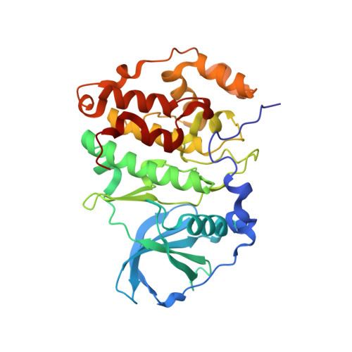
Crystal structure of the Rho-associated coiled-coil kinase 2 inhibitor belumosudil bound to CK2 alpha.

Brear, P.Hyvonen, M.

(2022) Acta Crystallogr F Struct Biol Commun 78: 348-353

  • DOI: https://doi.org/10.1107/S2053230X22008767
  • Primary Citation of Related Structures:  
    7Z39

  • PubMed Abstract: 

    The small molecule belumosudil was initially identified as a selective inhibitor of Rho-associated coiled-coil kinase 2 (ROCK2) and has recently been approved for the treatment of graft-versus-host disease. However, recent studies have shown that many of the phenotypes displayed upon treatment with belumosudil were due to CK2α inhibition. CK2α is in itself a very promising therapeutic target for a range of conditions and has recently been put forward as a potential treatment for COVID-19. Belumosudil presents a promising starting point for the development of future CK2α inhibitors as it provides a safe, potent and orally bioavailable scaffold. Therefore, several of the major hurdles in drug development have already been overcome. Here, the crystal structure of belumosudil bound to the ATP site of CK2α is presented. This crystal structure combined with modelling studies further elucidates how belumosudil could be developed into a selective and potent CK2α or ROCK2 inhibitor.


  • Organizational Affiliation

    Department of Biochemistry, University of Cambridge, 80 Tennis Court Road, Cambridge, United Kingdom.


Macromolecules
Find similar proteins by:  (by identity cutoff)  |  3D Structure
Entity ID: 1
MoleculeChains Sequence LengthOrganismDetailsImage
Casein kinase II subunit alpha328Homo sapiensMutation(s): 4 
Gene Names: CSNK2A1CK2A1
EC: 2.7.11.1
UniProt & NIH Common Fund Data Resources
Find proteins for P68400 (Homo sapiens)
Explore P68400 
Go to UniProtKB:  P68400
PHAROS:  P68400
GTEx:  ENSG00000101266 
Entity Groups  
Sequence Clusters30% Identity50% Identity70% Identity90% Identity95% Identity100% Identity
UniProt GroupP68400
Sequence Annotations
Expand
  • Reference Sequence
Experimental Data & Validation

Experimental Data

  • Method: X-RAY DIFFRACTION
  • Resolution: 1.60 Å
  • R-Value Free: 0.213 
  • R-Value Work: 0.164 
  • R-Value Observed: 0.166 
  • Space Group: P 1 21 1
Unit Cell:
Length ( Å )Angle ( ˚ )
a = 58.508α = 90
b = 45.753β = 112.47
c = 63.462γ = 90
Software Package:
Software NamePurpose
Aimlessdata scaling
REFMACrefinement
PDB_EXTRACTdata extraction
XDSdata reduction
PHASERphasing

Structure Validation

View Full Validation Report


Ligand Structure Quality Assessment 


Entry History & Funding Information

Deposition Data


Funding OrganizationLocationGrant Number
University of CambridgeUnited Kingdom--

Revision History  (Full details and data files)

  • Version 1.0: 2022-10-12
    Type: Initial release
  • Version 1.1: 2023-03-29
    Changes: Database references
  • Version 1.2: 2024-02-07
    Changes: Data collection, Refinement description