7Z37 | pdb_00007z37

Structure of the RAF1-HSP90-CDC37 complex (RHC-II)


Experimental Data Snapshot

  • Method: ELECTRON MICROSCOPY
  • Resolution: 3.67 Å
  • Aggregation State: PARTICLE 
  • Reconstruction Method: SINGLE PARTICLE 

wwPDB Validation 3D Report Full Report

Validation slider image for 7Z37

This is version 1.2 of the entry. See complete history

Literature

Structure of the RAF1-HSP90-CDC37 complex reveals the basis of RAF1 regulation.

Garcia-Alonso, S.Mesa, P.Ovejero, L.P.Aizpurua, G.Lechuga, C.G.Zarzuela, E.Santiveri, C.M.Sanclemente, M.Munoz, J.Musteanu, M.Campos-Olivas, R.Martinez-Torrecuadrada, J.Barbacid, M.Montoya, G.

(2022) Mol Cell 82: 3438-3452.e8

  • DOI: https://doi.org/10.1016/j.molcel.2022.08.012
  • Primary Citation Related Structures: 
    7Z37, 7Z38

  • PubMed Abstract: 

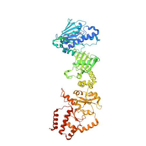
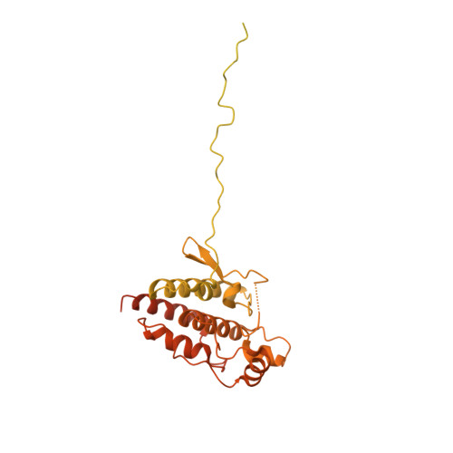
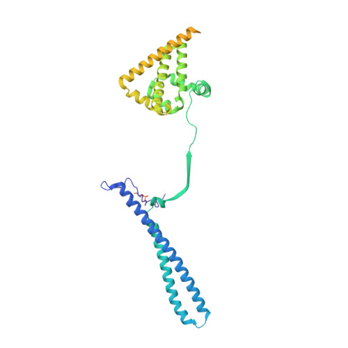
    RAF kinases are RAS-activated enzymes that initiate signaling through the MAPK cascade to control cellular proliferation, differentiation, and survival. Here, we describe the structure of the full-length RAF1 protein in complex with HSP90 and CDC37 obtained by cryoelectron microscopy. The reconstruction reveals a RAF1 kinase with an unfolded N-lobe separated from its C-lobe. The hydrophobic core of the N-lobe is trapped in the HSP90 dimer, while CDC37 wraps around the chaperone and interacts with the N- and C-lobes of the kinase. The structure indicates how CDC37 can discriminate between the different members of the RAF family. Our structural analysis also reveals that the folded RAF1 assembles with 14-3-3 dimers, suggesting that after folding RAF1 follows a similar activation as B-RAF. Finally, disruption of the interaction between CDC37 and the DFG segment of RAF1 unveils potential vulnerabilities in attempting the pharmacological degradation of RAF1 for therapeutic purposes.


  • Organizational Affiliation
    • Experimental Oncology Group, Molecular Oncology Programme, Centro Nacional de Investigaciones Oncológicas (CNIO), Madrid 28029, Spain; Centro de Investigación Biomédica en Red de Cáncer (CIBERONC), Instituto de Salud Carlos III, 28029 Madrid, Spain.

Macromolecule Content 

  • Total Structure Weight: 290.7 kDa 
  • Atom Count: 14,256 
  • Modeled Residue Count: 1,740 
  • Deposited Residue Count: 2,517 
  • Unique protein chains: 3

Macromolecules

Find similar proteins by:|  3D Structure
Entity ID: 1
MoleculeChains  Sequence LengthOrganismDetailsImage
Heat shock protein HSP 90-betaA [auth AP1],
B [auth BP1]
732Homo sapiensMutation(s): 0 
Gene Names: HSP90AB1HSP90BHSPC2HSPCB
UniProt & NIH Common Fund Data Resources
Find proteins for P08238 (Homo sapiens)
Explore P08238 
Go to UniProtKB:  P08238
PHAROS:  P08238
GTEx:  ENSG00000096384 
Entity Groups
Sequence Clusters30% Identity50% Identity70% Identity90% Identity95% Identity100% Identity
UniProt GroupP08238
Sequence Annotations
Expand
Reference Sequence
Find similar proteins by:|  3D Structure
Entity ID: 2
MoleculeChains  Sequence LengthOrganismDetailsImage
RAF proto-oncogene serine/threonine-protein kinaseC [auth CP1]659Homo sapiensMutation(s): 0 
Gene Names: RAF1RAF
EC: 2.7.11.1
UniProt & NIH Common Fund Data Resources
Find proteins for P04049 (Homo sapiens)
Explore P04049 
Go to UniProtKB:  P04049
PHAROS:  P04049
GTEx:  ENSG00000132155 
Entity Groups
Sequence Clusters30% Identity50% Identity70% Identity90% Identity95% Identity100% Identity
UniProt GroupP04049
Sequence Annotations
Expand
Reference Sequence
Find similar proteins by:|  3D Structure
Entity ID: 3
MoleculeChains  Sequence LengthOrganismDetailsImage
Hsp90 co-chaperone Cdc37D [auth DP1]394Homo sapiensMutation(s): 0 
Gene Names: CDC37CDC37A
UniProt & NIH Common Fund Data Resources
Find proteins for Q16543 (Homo sapiens)
Explore Q16543 
Go to UniProtKB:  Q16543
PHAROS:  Q16543
GTEx:  ENSG00000105401 
Entity Groups
Sequence Clusters30% Identity50% Identity70% Identity90% Identity95% Identity100% Identity
UniProt GroupQ16543
Sequence Annotations
Expand
Reference Sequence

Small Molecules

Modified Residues  1 Unique
IDChains TypeFormula2D DiagramParent
SEP
Query on SEP
D [auth DP1]L-PEPTIDE LINKINGC3 H8 N O6 PSER

Experimental Data & Validation

Experimental Data

  • Method: ELECTRON MICROSCOPY
  • Resolution: 3.67 Å
  • Aggregation State: PARTICLE 
  • Reconstruction Method: SINGLE PARTICLE 
EM Software:
TaskSoftware PackageVersion
RECONSTRUCTIONRELION3.1.3
MODEL REFINEMENTPHENIX
MODEL REFINEMENTISOLDE

Structure Validation

View Full Validation Report


Entry History 

& Funding Information

Deposition Data


Funding OrganizationLocationGrant Number
Novo Nordisk FoundationDenmarkNNF14CC0001
Novo Nordisk FoundationDenmarkNNF0024386
Novo Nordisk FoundationDenmarkNNF17SA0030214
Novo Nordisk FoundationDenmarkNNF18OC0055061

Revision History  (Full details and data files)

  • Version 1.0: 2022-09-14
    Type: Initial release
  • Version 1.1: 2022-09-28
    Changes: Database references
  • Version 1.2: 2024-11-20
    Changes: Data collection, Structure summary