In Vivo Photocontrol of Microtubule Dynamics and Integrity, Migration and Mitosis, by the Potent GFP-Imaging-Compatible Photoswitchable Reagents SBTubA4P and SBTub2M.
Gao, L., Meiring, J.C.M., Varady, A., Ruider, I.E., Heise, C., Wranik, M., Velasco, C.D., Taylor, J.A., Terni, B., Weinert, T., Standfuss, J., Cabernard, C.C., Llobet, A., Steinmetz, M.O., Bausch, A.R., Distel, M., Thorn-Seshold, J., Akhmanova, A., Thorn-Seshold, O.(2022) J Am Chem Soc 144: 5614-5628
- PubMed: 35290733 Search on PubMedSearch on PubMed Central
- DOI: https://doi.org/10.1021/jacs.2c01020
- Primary Citation Related Structures:
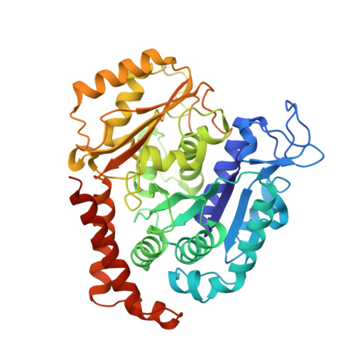
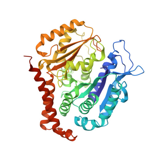
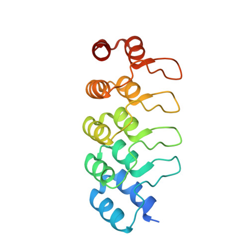
7Z01, 7Z02 - PubMed Abstract:
Photoswitchable reagents are powerful tools for high-precision studies in cell biology. When these reagents are globally administered yet locally photoactivated in two-dimensional (2D) cell cultures, they can exert micron- and millisecond-scale biological control. This gives them great potential for use in biologically more relevant three-dimensional (3D) models and in vivo , particularly for studying systems with inherent spatiotemporal complexity, such as the cytoskeleton. However, due to a combination of photoswitch isomerization under typical imaging conditions, metabolic liabilities, and insufficient water solubility at effective concentrations, the in vivo potential of photoswitchable reagents addressing cytosolic protein targets remains largely unrealized. Here, we optimized the potency and solubility of metabolically stable, druglike colchicinoid microtubule inhibitors based on the styrylbenzothiazole (SBT) scaffold that are nonresponsive to typical fluorescent protein imaging wavelengths and so enable multichannel imaging studies. We applied these reagents both to 3D organoids and tissue explants and to classic model organisms (zebrafish, clawed frog) in one- and two-protein imaging experiments, in which spatiotemporally localized illuminations allowed them to photocontrol microtubule dynamics, network architecture, and microtubule-dependent processes in vivo with cellular precision and second-level resolution. These nanomolar, in vivo capable photoswitchable reagents should open up new dimensions for high-precision cytoskeleton research in cargo transport, cell motility, cell division, and development. More broadly, their design can also inspire similarly capable optical reagents for a range of cytosolic protein targets, thus bringing in vivo photopharmacology one step closer to general realization.
- Department of Pharmacy, Ludwig-Maximilians University of Munich, Munich 81377, Germany.
Organizational Affiliation: