7YZ9

Structure of catalytic domain of Rv1625c bound to nanobody NB4


Experimental Data Snapshot

  • Method: X-RAY DIFFRACTION
  • Resolution: 1.97 Å
  • R-Value Free: 0.244 
  • R-Value Work: 0.206 
  • R-Value Observed: 0.208 

wwPDB Validation   3D Report Full Report


Ligand Structure Quality Assessment 


This is version 1.1 of the entry. See complete history


Literature

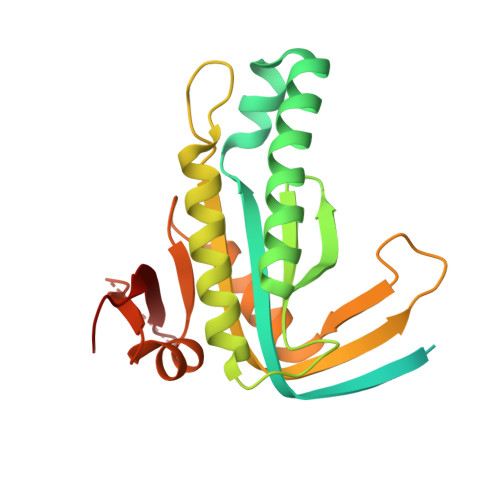
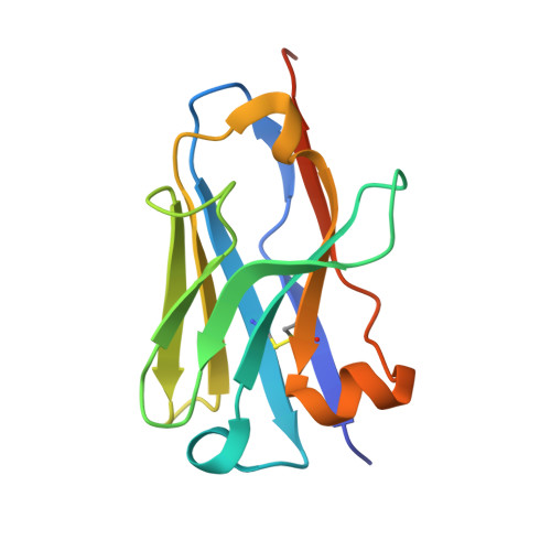
Structure of Mycobacterium tuberculosis Cya, an evolutionary ancestor of the mammalian membrane adenylyl cyclases.

Mehta, V.Khanppnavar, B.Schuster, D.Kantarci, I.Vercellino, I.Kosturanova, A.Iype, T.Stefanic, S.Picotti, P.Korkhov, V.M.

(2022) Elife 11

  • DOI: https://doi.org/10.7554/eLife.77032
  • Primary Citation of Related Structures:  
    7YZ9, 7YZI, 7YZK

  • PubMed Abstract: 

    Mycobacterium tuberculosis adenylyl cyclase (AC) Rv1625c/Cya is an evolutionary ancestor of the mammalian membrane ACs and a model system for studies of their structure and function. Although the vital role of ACs in cellular signalling is well established, the function of their transmembrane (TM) regions remains unknown. Here, we describe the cryo-EM structure of Cya bound to a stabilizing nanobody at 3.6 Å resolution. The TM helices 1-5 form a structurally conserved domain that facilitates the assembly of the helical and catalytic domains. The TM region contains discrete pockets accessible from the extracellular and cytosolic side of the membrane. Neutralization of the negatively charged extracellular pocket Ex1 destabilizes the cytosolic helical domain and reduces the catalytic activity of the enzyme. The TM domain acts as a functional component of Cya, guiding the assembly of the catalytic domain and providing the means for direct regulation of catalytic activity in response to extracellular ligands.


  • Organizational Affiliation

    Laboratory of Biomolecular Research, Division of Biology and Chemistry, Paul Scherrer Institute, Villigen, Switzerland.


Macromolecules
Find similar proteins by:  (by identity cutoff)  |  3D Structure
Entity ID: 1
MoleculeChains Sequence LengthOrganismDetailsImage
Adenylate cyclase254Mycobacterium tuberculosis H37RvMutation(s): 0 
Gene Names: cyaRv1625cMTCY01B2.17c
EC: 4.6.1.1
UniProt
Find proteins for P9WQ35 (Mycobacterium tuberculosis (strain ATCC 25618 / H37Rv))
Explore P9WQ35 
Go to UniProtKB:  P9WQ35
Entity Groups  
Sequence Clusters30% Identity50% Identity70% Identity90% Identity95% Identity100% Identity
UniProt GroupP9WQ35
Sequence Annotations
Expand
  • Reference Sequence
Find similar proteins by:  (by identity cutoff)  |  3D Structure
Entity ID: 2
MoleculeChains Sequence LengthOrganismDetailsImage
nanobody NB4128Vicugna pacosMutation(s): 0 
Entity Groups  
Sequence Clusters30% Identity50% Identity70% Identity90% Identity95% Identity100% Identity
Sequence Annotations
Expand
  • Reference Sequence
Experimental Data & Validation

Experimental Data

  • Method: X-RAY DIFFRACTION
  • Resolution: 1.97 Å
  • R-Value Free: 0.244 
  • R-Value Work: 0.206 
  • R-Value Observed: 0.208 
  • Space Group: P 65 2 2
Unit Cell:
Length ( Å )Angle ( ˚ )
a = 94.793α = 90
b = 94.793β = 90
c = 119.655γ = 120
Software Package:
Software NamePurpose
PHENIXrefinement
PHENIXrefinement
iMOSFLMdata reduction
XDSdata scaling
PHENIXphasing

Structure Validation

View Full Validation Report


Ligand Structure Quality Assessment 


Entry History & Funding Information

Deposition Data


Funding OrganizationLocationGrant Number
Swiss National Science FoundationSwitzerland150665
Swiss National Science FoundationSwitzerland176992
Swiss National Science FoundationSwitzerland184951

Revision History  (Full details and data files)

  • Version 1.0: 2022-08-31
    Type: Initial release
  • Version 1.1: 2024-01-31
    Changes: Data collection, Refinement description