7YXJ

Drosophila melanogaster JMJD7 (dmJMJD7) in complex with Mn and 2,4-PDCA


Experimental Data Snapshot

  • Method: X-RAY DIFFRACTION
  • Resolution: 2.45 Å
  • R-Value Free: 0.230 
  • R-Value Work: 0.206 
  • R-Value Observed: 0.207 

wwPDB Validation   3D Report Full Report


Ligand Structure Quality Assessment 


This is version 1.2 of the entry. See complete history


Literature

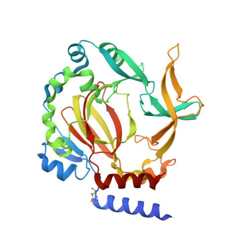
Conservation of the unusual dimeric JmjC fold of JMJD7 from Drosophila melanogaster to humans.

Chowdhury, R.Abboud, M.I.Wiley, J.Tumber, A.Markolovic, S.Schofield, C.J.

(2022) Sci Rep 12: 6065-6065

  • DOI: https://doi.org/10.1038/s41598-022-10028-y
  • Primary Citation of Related Structures:  
    7YXG, 7YXH, 7YXI, 7YXJ, 7YXK, 7YXL

  • PubMed Abstract: 

    The JmjC family of 2-oxoglutarate dependent oxygenases catalyse a range of hydroxylation and demethylation reactions in humans and other animals. Jumonji domain-containing 7 (JMJD7) is a JmjC (3S)-lysyl-hydroxylase that catalyses the modification of Developmentally Regulated GTP Binding Proteins 1 and 2 (DRG1 and 2); JMJD7 has also been reported to have histone endopeptidase activity. Here we report biophysical and biochemical studies on JMJD7 from Drosophila melanogaster (dmJMJD7). Notably, crystallographic analyses reveal that the unusual dimerization mode of JMJD7, which involves interactions between both the N- and C-terminal regions of both dmJMJD7 monomers and disulfide formation, is conserved in human JMJD7 (hsJMJD7). The results further support the assignment of JMJD7 as a lysyl hydroxylase and will help enable the development of selective inhibitors for it and other JmjC oxygenases.


  • Organizational Affiliation

    Chemistry Research Laboratory, Department of Chemistry and the Ineos Oxford Institute for Antimicrobial Research, University of Oxford, Mansfield Road, Oxford, OX1 3TA, UK.


Macromolecules
Find similar proteins by:  (by identity cutoff)  |  3D Structure
Entity ID: 1
MoleculeChains Sequence LengthOrganismDetailsImage
GH14974p
A, B, C, D
322Drosophila melanogasterMutation(s): 0 
Gene Names: JMJD7Dmel\CG10133CG10133Dmel_CG10133
EC: 1.14.11
UniProt
Find proteins for Q9VU77 (Drosophila melanogaster)
Explore Q9VU77 
Go to UniProtKB:  Q9VU77
Entity Groups  
Sequence Clusters30% Identity50% Identity70% Identity90% Identity95% Identity100% Identity
UniProt GroupQ9VU77
Sequence Annotations
Expand
  • Reference Sequence
Small Molecules
Ligands 4 Unique
IDChains Name / Formula / InChI Key2D Diagram3D Interactions
PD2 (Subject of Investigation/LOI)
Query on PD2

Download Ideal Coordinates CCD File 
K [auth A],
Q [auth B],
T [auth C],
Z [auth D]
PYRIDINE-2,4-DICARBOXYLIC ACID
C7 H5 N O4
MJIVRKPEXXHNJT-UHFFFAOYSA-N
PEG
Query on PEG

Download Ideal Coordinates CCD File 
F [auth A]DI(HYDROXYETHYL)ETHER
C4 H10 O3
MTHSVFCYNBDYFN-UHFFFAOYSA-N
EDO
Query on EDO

Download Ideal Coordinates CCD File 
G [auth A]
H [auth A]
I [auth A]
J [auth A]
M [auth B]
G [auth A],
H [auth A],
I [auth A],
J [auth A],
M [auth B],
N [auth B],
O [auth B],
P [auth B],
S [auth C],
V [auth D],
W [auth D],
X [auth D],
Y [auth D]
1,2-ETHANEDIOL
C2 H6 O2
LYCAIKOWRPUZTN-UHFFFAOYSA-N
MN
Query on MN

Download Ideal Coordinates CCD File 
E [auth A],
L [auth B],
R [auth C],
U [auth D]
MANGANESE (II) ION
Mn
WAEMQWOKJMHJLA-UHFFFAOYSA-N
Modified Residues  1 Unique
IDChains TypeFormula2D DiagramParent
CSO
Query on CSO
A, B, C, D
L-PEPTIDE LINKINGC3 H7 N O3 SCYS
Experimental Data & Validation

Experimental Data

  • Method: X-RAY DIFFRACTION
  • Resolution: 2.45 Å
  • R-Value Free: 0.230 
  • R-Value Work: 0.206 
  • R-Value Observed: 0.207 
  • Space Group: P 1 21 1
Unit Cell:
Length ( Å )Angle ( ˚ )
a = 53.557α = 90
b = 65.762β = 97.78
c = 206.957γ = 90
Software Package:
Software NamePurpose
PHENIXrefinement
SCALAdata scaling
PDB_EXTRACTdata extraction
XDSdata reduction
PHASERphasing

Structure Validation

View Full Validation Report


Ligand Structure Quality Assessment 


Entry History & Funding Information

Deposition Data


Funding OrganizationLocationGrant Number
Wellcome TrustUnited Kingdom106244/Z/14/Z

Revision History  (Full details and data files)

  • Version 1.0: 2022-04-27
    Type: Initial release
  • Version 1.1: 2022-05-04
    Changes: Refinement description
  • Version 1.2: 2024-01-31
    Changes: Data collection, Refinement description