7YUI

Crystal structure of HOIL-1L(195-423) in complex with the linear tetra-ubiquitin


Experimental Data Snapshot

  • Method: X-RAY DIFFRACTION
  • Resolution: 2.60 Å
  • R-Value Free: 0.258 
  • R-Value Work: 0.203 
  • R-Value Observed: 0.205 

wwPDB Validation   3D Report Full Report


This is version 1.1 of the entry. See complete history


Literature

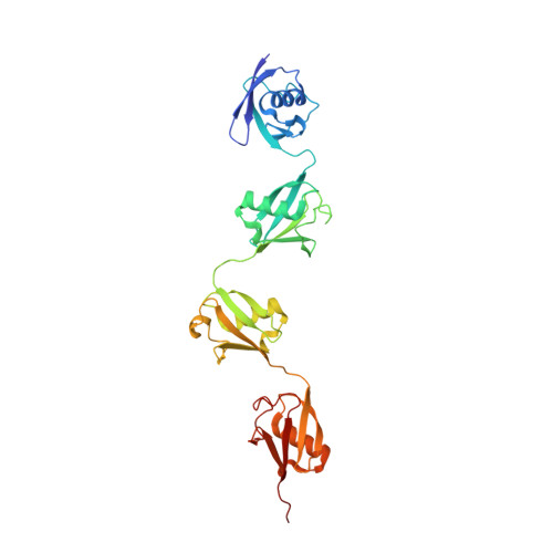
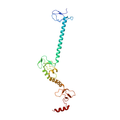
Mechanistic insights into the enzymatic activity of E3 ligase HOIL-1L and its regulation by the linear ubiquitin chain binding.

Xu, X.Wang, Y.Zhang, Y.Wang, Y.Yin, Y.Peng, C.Gong, X.Li, M.Zhang, Y.Zhang, M.Tang, Y.Zhou, X.Liu, H.Pan, L.

(2023) Sci Adv 9: eadi4599-eadi4599

  • DOI: https://doi.org/10.1126/sciadv.adi4599
  • Primary Citation of Related Structures:  
    7YUI, 7YUJ

  • PubMed Abstract: 

    Heme-oxidized IRP2 ubiquitin ligase 1 (HOIL-1L) serves as a unique E3 ligase to catalyze the mono-ubiquitination of relevant protein or sugar substrates and plays vital roles in numerous cellular processes in mammals. However, the molecular mechanism underpinning the E3 activity of HOIL-1L and the related regulatory mechanism remain elusive. Here, we report the crystal structure of the catalytic core region of HOIL-1L and unveil the key catalytic triad residues of HOIL-1L. Moreover, we discover that HOIL-1L contains two distinct linear di-ubiquitin binding sites that can synergistically bind to linear tetra-ubiquitin, and the binding of HOIL-1L with linear tetra-ubiquitin can promote its E3 activity. The determined HOIL-1L/linear tetra-ubiquitin complex structure not only elucidates the detailed binding mechanism of HOIL-1L with linear tetra-ubiquitin but also uncovers a unique allosteric ubiquitin-binding site for the activation of HOIL-1L. In all, our findings provide mechanistic insights into the E3 activity of HOIL-1L and its regulation by the linear ubiquitin chain binding.


  • Organizational Affiliation

    School of Chemistry and Materials Science, Hangzhou Institute for Advanced Study, University of Chinese Academy of Sciences, 1 Sub-lane Xiangshan, Hangzhou 310024, China.


Macromolecules
Find similar proteins by:  (by identity cutoff)  |  3D Structure
Entity ID: 1
MoleculeChains Sequence LengthOrganismDetailsImage
Polyubiquitin-C308Homo sapiensMutation(s): 0 
Gene Names: UBC
UniProt & NIH Common Fund Data Resources
Find proteins for P0CG48 (Homo sapiens)
Explore P0CG48 
Go to UniProtKB:  P0CG48
PHAROS:  P0CG48
GTEx:  ENSG00000150991 
Entity Groups  
Sequence Clusters30% Identity50% Identity70% Identity90% Identity95% Identity100% Identity
UniProt GroupP0CG48
Sequence Annotations
Expand
  • Reference Sequence
Find similar proteins by:  (by identity cutoff)  |  3D Structure
Entity ID: 2
MoleculeChains Sequence LengthOrganismDetailsImage
RanBP-type and C3HC4-type zinc finger-containing protein 1234Homo sapiensMutation(s): 0 
Gene Names: RBCK1
EC: 2.3.2.31
UniProt & NIH Common Fund Data Resources
Find proteins for Q9BYM8 (Homo sapiens)
Explore Q9BYM8 
Go to UniProtKB:  Q9BYM8
PHAROS:  Q9BYM8
GTEx:  ENSG00000125826 
Entity Groups  
Sequence Clusters30% Identity50% Identity70% Identity90% Identity95% Identity100% Identity
UniProt GroupQ9BYM8
Sequence Annotations
Expand
  • Reference Sequence
Experimental Data & Validation

Experimental Data

  • Method: X-RAY DIFFRACTION
  • Resolution: 2.60 Å
  • R-Value Free: 0.258 
  • R-Value Work: 0.203 
  • R-Value Observed: 0.205 
  • Space Group: P 21 21 21
Unit Cell:
Length ( Å )Angle ( ˚ )
a = 58.5α = 90
b = 90.304β = 90
c = 117.439γ = 90
Software Package:
Software NamePurpose
PHENIXrefinement
PDB_EXTRACTdata extraction
XDSdata reduction
Aimlessdata scaling
PHASERphasing

Structure Validation

View Full Validation Report


Entry History & Funding Information

Deposition Data


Funding OrganizationLocationGrant Number
National Natural Science Foundation of China (NSFC)China21621002

Revision History  (Full details and data files)

  • Version 1.0: 2023-08-30
    Type: Initial release
  • Version 1.1: 2024-03-13
    Changes: Database references, Structure summary