A blocking monoclonal antibody reveals dimerization of intracellular domains of ALK2 associated with genetic disorders.
Katagiri, T., Tsukamoto, S., Kuratani, M., Tsuji, S., Nakamura, K., Ohte, S., Kawaguchi, Y., Takaishi, K.(2023) Nat Commun 14: 2960-2960
- PubMed: 37231012 Search on PubMedSearch on PubMed Central
- DOI: https://doi.org/10.1038/s41467-023-38746-5
- Primary Citation Related Structures:
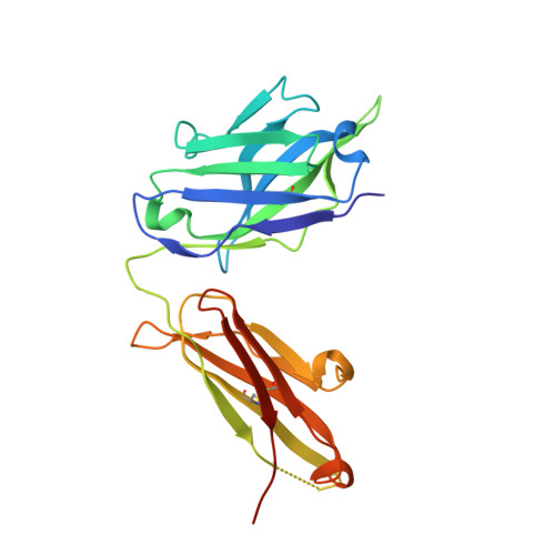
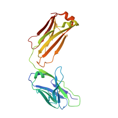
7YRU - PubMed Abstract:
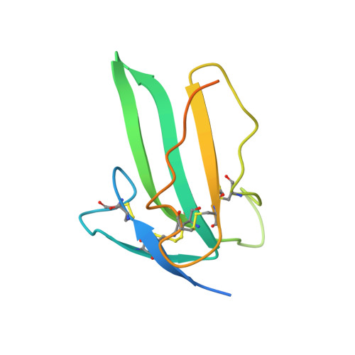
Mutations in activin receptor-like kinase 2 (ALK2) can cause the pathological osteogenic signaling seen in some patients with fibrodysplasia ossificans progressiva and other conditions such as diffuse intrinsic pontine glioma. Here, we report that intracellular domain of wild-type ALK2 readily dimerizes in response to BMP7 binding to drive osteogenic signaling. This osteogenic signaling is pathologically triggered by heterotetramers of type II receptor kinases and ALK2 mutant forms, which form intracellular domain dimers in response to activin A binding. We develop a blocking monoclonal antibody, Rm0443, that can suppress ALK2 signaling. We solve the crystal structure of the ALK2 extracellular domain complex with a Fab fragment of Rm0443 and show that Rm0443 induces dimerization of ALK2 extracellular domains in a back-to-back orientation on the cell membrane by binding the residues H64 and F63 on opposite faces of the ligand-binding site. Rm0443 could prevent heterotopic ossification in a mouse model of fibrodysplasia ossificans progressiva that carries the human R206H pathogenic mutant.
- Division of Biomedical Sciences, Research Center for Genomic Medicine, Saitama Medical University, 1397-1 Yamane, Hidaka-shi, Saitama, 350-1241, Japan. katagiri@saitama-med.ac.jp.
Organizational Affiliation: