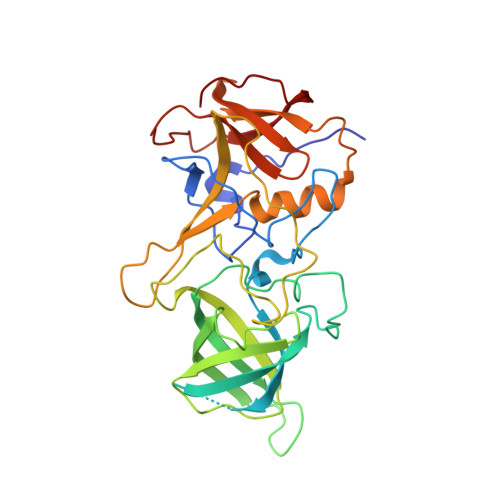
Functional and structural characterization of Norovirus GII.6 in recognizing histo-blood group antigens.
Cong, X., Li, H.B., Sun, X.M., Qi, J.X., Zhang, Q., Duan, Z.J., Xu, Y., Liu, W.L.(2023) Virol Sin 38: 56-65
- PubMed: 36216242 Search on PubMedSearch on PubMed Central
- DOI: https://doi.org/10.1016/j.virs.2022.09.010
- Primary Citation Related Structures:
7YQB, 7YQG - PubMed Abstract:
Noroviruses (NoVs) are the primary cause of acute gastroenteritis worldwide. Histo-blood group antigens (HBGAs) are receptors or attachment factors that affect the prevalence and host susceptibility of NoVs. GII.6 NoV is one of the predominant genotypes in humans, which recognizes the type ABO secretor of HBGAs. However, the structural basis of GII.6 NoV's interaction with HBGAs receptors remains elusive. In this study, we investigated the binding features of the GII.6 strain to HBGAs using saliva- and glycan-ELISA assays and characterized the molecular basis of the GII.6 virus that recognizes H disaccharide. We showed that the GII.6 P domain recognized some A and O secretor's saliva samples, most B secretor's saliva samples, and H disaccharide antigen, but did not bind non-secretors' saliva. Further, we determined the crystal structures of GII.6 and its complex with H disaccharides at 1.7 Å, revealing that the P domain of GII.6 shares the conventional binding interface and mode of GII HBGAs. Single residue mutations at the GII.6-H binding sites could inhibit the binding of GII.6 to HBGAs, demonstrating that the interaction residues were crucial in maintaining NoV-glycan integrity. Finally, structural and sequence analyses showed that the major residues of the GII.6-H interaction were conserved among NoVs in the GII genogroup. Taken together, our study characterized the functional and structural features of GII.6 that allow it to interact with HBGAs, and shed light on NoV evolution, epidemiology, and anti-viral drug development.
- Department of Neurosurgery, Shenzhen Second People's Hospital/the First Affiliated Hospital of Shenzhen University Health Science Center, Shenzhen, 518035, China; The Center for Medical Genetics & Molecular Diagnosis, Shenzhen Second People's Hospital/the First Affiliated Hospital of Shenzhen University Health Science Center, Shenzhen, 518035, China.
Organizational Affiliation: