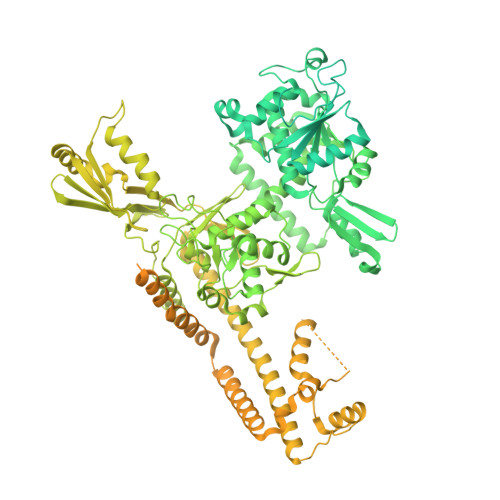
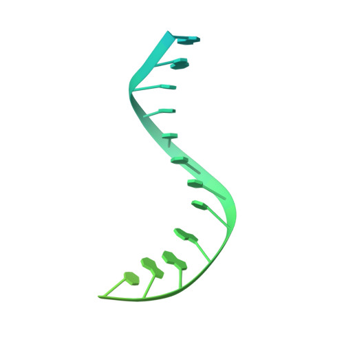
ERK2-topoisomerase II regulatory axis is important for gene activation in immediate early genes.
Bunch, H., Kim, D., Naganuma, M., Nakagawa, R., Cong, A., Jeong, J., Ehara, H., Vu, H., Chang, J.H., Schellenberg, M.J., Sekine, S.I.(2023) Nat Commun 14: 8341-8341
- PubMed: 38097570 Search on PubMedSearch on PubMed Central
- DOI: https://doi.org/10.1038/s41467-023-44089-y
- Primary Citation Related Structures:
7YQ8 - PubMed Abstract:
The function of the mitogen-activated protein kinase signaling pathway is required for the activation of immediate early genes (IEGs), including EGR1 and FOS, for cell growth and proliferation. Recent studies have identified topoisomerase II (TOP2) as one of the important regulators of the transcriptional activation of IEGs. However, the mechanism underlying transcriptional regulation involving TOP2 in IEG activation has remained unknown. Here, we demonstrate that ERK2, but not ERK1, is important for IEG transcriptional activation and report a critical ELK1 binding sequence for ERK2 function at the EGR1 gene. Our data indicate that both ERK1 and ERK2 extensively phosphorylate the C-terminal domain of TOP2B at mutual and distinctive residues. Although both ERK1 and ERK2 enhance the catalytic rate of TOP2B required to relax positive DNA supercoiling, ERK2 delays TOP2B catalysis of negative DNA supercoiling. In addition, ERK1 may relax DNA supercoiling by itself. ERK2 catalytic inhibition or knock-down interferes with transcription and deregulates TOP2B in IEGs. Furthermore, we present the first cryo-EM structure of the human cell-purified TOP2B and etoposide together with the EGR1 transcriptional start site (-30 to +20) that has the strongest affinity to TOP2B within -423 to +332. The structure shows TOP2B-mediated breakage and dramatic bending of the DNA. Transcription is activated by etoposide, while it is inhibited by ICRF193 at EGR1 and FOS, suggesting that TOP2B-mediated DNA break to favor transcriptional activation. Taken together, this study suggests that activated ERK2 phosphorylates TOP2B to regulate TOP2-DNA interactions and favor transcriptional activation in IEGs. We propose that TOP2B association, catalysis, and dissociation on its substrate DNA are important processes for regulating transcription and that ERK2-mediated TOP2B phosphorylation may be key for the catalysis and dissociation steps.
- Department of Applied Biosciences, Kyungpook National University, Daegu, 41566, Republic of Korea. heeyounbunch@gmail.com.
Organizational Affiliation: