7YP3

Crystal structure of elaiophylin glycosyltransferase in complex with elaiophylin


Experimental Data Snapshot

  • Method: X-RAY DIFFRACTION
  • Resolution: 2.10 Å
  • R-Value Free: 0.217 
  • R-Value Work: 0.188 
  • R-Value Observed: 0.190 

wwPDB Validation   3D Report Full Report


Ligand Structure Quality Assessment 


This is version 1.1 of the entry. See complete history


Literature

Substrate-induced dimerization of elaiophylin glycosyltransferase reveals a novel self-activating form of glycosyltransferase for symmetric glycosylation.

Xu, T.Gan, Q.Liu, Q.Chen, R.Zhen, X.Zhang, C.Liu, J.

(2022) Acta Crystallogr D Struct Biol 78: 1235-1248

  • DOI: https://doi.org/10.1107/S2059798322008658
  • Primary Citation of Related Structures:  
    7YP3, 7YP4, 7YP5, 7YP6

  • PubMed Abstract: 

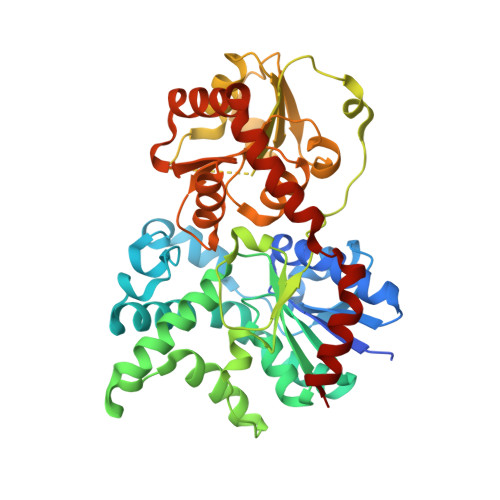
    Elaiophylin (Ela), a unique 16-membered symmetric macrodiolide antibiotic, displays broad biological activity. Two rare 2-deoxy-L-fucose moieties at the ends of Ela are critical for its activity. Previously, elaiophylin glycosyltransferase (ElaGT) was identified as the enzyme that is responsible for the symmetric glycosylation of Ela, acting as a potential enzymatic tool for enhancing the diversity and activity of Ela. However, a symmetric catalytic mechanism has never been reported for a glycosyltransferase (GT). To explore the catalytic mechanism, the structure of ElaGT was determined in four forms: the apo form and Ela-bound, thymidine diphosphate-bound and uridine diphosphate-bound forms. In the Ela-bound structure, two ElaGTs form a `face-to-face' C2-symmetric homodimer with a continuous acceptor-binding pocket, allowing a molecule of Ela to shuffle through. Interestingly, this dimer interface resembles that of the activator-dependent GT EryCIII with its activator EryCII. Sequence analysis also indicates that ElaGT belongs to the activator-dependent GT family, but no putative activator has been identified in the Ela gene cluster. It was then found that the ElaGT homodimer may utilize this `face-to-face' arrangement to stabilize the Ela-binding loops on the interface and to simultaneously allosterically regulate the catalytic center. Therefore, these structures present a novel self-activating model for symmetric sugar transfer in the GT family and a new potential regulation site for substrate specificity.


  • Organizational Affiliation

    State Key Laboratory of Respiratory Disease, Guangzhou Institutes of Biomedicine and Health, Chinese Academy of Sciences, Guangzhou 510530, People's Republic of China.


Macromolecules
Find similar proteins by:  (by identity cutoff)  |  3D Structure
Entity ID: 1
MoleculeChains Sequence LengthOrganismDetailsImage
Glycosyltransferase
A, B, C, D, E
A, B, C, D, E, F, G
437Streptomyces sp. SCSIO 01934Mutation(s): 0 
UniProt
Find proteins for E5L4T5 (Streptomyces sp. SCSIO 01934)
Explore E5L4T5 
Go to UniProtKB:  E5L4T5
Entity Groups  
Sequence Clusters30% Identity50% Identity70% Identity90% Identity95% Identity100% Identity
UniProt GroupE5L4T5
Sequence Annotations
Expand
  • Reference Sequence
Small Molecules
Ligands 3 Unique
IDChains Name / Formula / InChI Key2D Diagram3D Interactions
ELO (Subject of Investigation/LOI)
Query on ELO

Download Ideal Coordinates CCD File 
M [auth B],
R [auth C],
T [auth D],
W [auth E],
Z [auth F]
Elaiophylin
C54 H88 O18
OSERMIPXNLXAPD-MJMYBOKFSA-N
GOL
Query on GOL

Download Ideal Coordinates CCD File 
DA [auth F]
EA [auth F]
FA [auth F]
JA [auth G]
K [auth A]
DA [auth F],
EA [auth F],
FA [auth F],
JA [auth G],
K [auth A],
KA [auth G],
L [auth A],
P [auth B],
Q [auth B],
S [auth C],
Y [auth E]
GLYCEROL
C3 H8 O3
PEDCQBHIVMGVHV-UHFFFAOYSA-N
ACT
Query on ACT

Download Ideal Coordinates CCD File 
AA [auth F]
BA [auth F]
CA [auth F]
GA [auth G]
H [auth A]
AA [auth F],
BA [auth F],
CA [auth F],
GA [auth G],
H [auth A],
HA [auth G],
I [auth A],
IA [auth G],
J [auth A],
N [auth B],
O [auth B],
U [auth D],
V [auth D],
X [auth E]
ACETATE ION
C2 H3 O2
QTBSBXVTEAMEQO-UHFFFAOYSA-M
Experimental Data & Validation

Experimental Data

  • Method: X-RAY DIFFRACTION
  • Resolution: 2.10 Å
  • R-Value Free: 0.217 
  • R-Value Work: 0.188 
  • R-Value Observed: 0.190 
  • Space Group: C 1 2 1
Unit Cell:
Length ( Å )Angle ( ˚ )
a = 228.098α = 90
b = 98.134β = 103.26
c = 176.506γ = 90
Software Package:
Software NamePurpose
REFMACrefinement
Aimlessdata scaling
PDB_EXTRACTdata extraction
DIALSdata reduction
PHASERphasing

Structure Validation

View Full Validation Report


Ligand Structure Quality Assessment 


Entry History & Funding Information

Deposition Data


Funding OrganizationLocationGrant Number
National Natural Science Foundation of China (NSFC)China31170708

Revision History  (Full details and data files)

  • Version 1.0: 2022-11-02
    Type: Initial release
  • Version 1.1: 2023-11-29
    Changes: Data collection, Refinement description