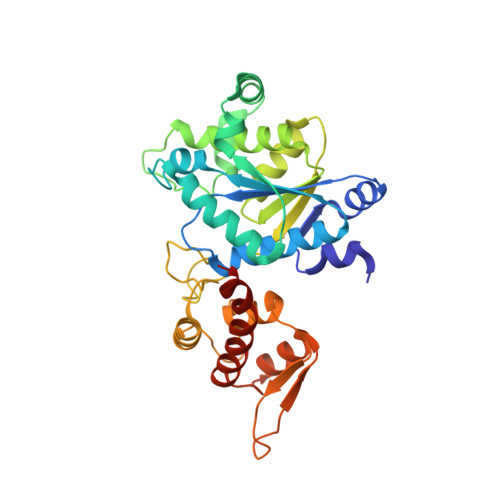
7YNW

Crystal structure of O-(2-nitrobenzyl)-L-tyrosine-tRNA sythetase in complex with O-(2-nitrobenzyl)-L-tyrosine


Experimental Data Snapshot

  • Method: X-RAY DIFFRACTION
  • Resolution: 2.79 Å
  • R-Value Free: 0.297 
  • R-Value Work: 0.260 
  • R-Value Observed: 0.262 

wwPDB Validation   3D Report Full Report


Ligand Structure Quality Assessment 


This is version 2.1 of the entry. See complete history


Literature

Crystal Structure of an Archaeal Tyrosyl-tRNA Synthetase Bound to Photocaged L-Tyrosine and Its Potential Application to Time-Resolved X-ray Crystallography.

Hosaka, T.Katsura, K.Ishizuka-Katsura, Y.Hanada, K.Ito, K.Tomabechi, Y.Inoue, M.Akasaka, R.Takemoto, C.Shirouzu, M.

(2022) Int J Mol Sci 23

  • DOI: https://doi.org/10.3390/ijms231810399
  • Primary Citation of Related Structures:  
    7YNU, 7YNV, 7YNW

  • PubMed Abstract: 

    Genetically encoded caged amino acids can be used to control the dynamics of protein activities and cellular localization in response to external cues. In the present study, we revealed the structural basis for the recognition of O -(2-nitrobenzyl)-L-tyrosine ( o NBTyr) by its specific variant of Methanocaldococcus jannaschii tyrosyl-tRNA synthetase ( o NBTyrRS), and then demonstrated its potential availability for time-resolved X-ray crystallography. The substrate-bound crystal structure of o NBTyrRS at a 2.79 Å resolution indicated that the replacement of tyrosine and leucine at positions 32 and 65 by glycine (Tyr32Gly and Leu65Gly, respectively) and Asp158Ser created sufficient space for entry of the bulky substitute into the amino acid binding pocket, while Glu in place of Leu162 formed a hydrogen bond with the nitro moiety of o NBTyr. We also produced an o NBTyr-containing lysozyme through a cell-free protein synthesis system derived from the Escherichia coli B95. ΔA strain with the UAG codon reassigned to the nonnatural amino acid. Another crystallographic study of the caged protein showed that the site-specifically incorporated o NBTyr was degraded to tyrosine by light irradiation of the crystals. Thus, cell-free protein synthesis of caged proteins with o NBTyr could facilitate time-resolved structural analysis of proteins, including medically important membrane proteins.


  • Organizational Affiliation

    Laboratory for Protein Functional and Structural Biology, RIKEN Center for Biosystems Dynamics Research, 1-7-22 Suehiro-cho, Tsurumi-ku, Yokohama 230-0045, Kanagawa, Japan.


Macromolecules
Find similar proteins by:  (by identity cutoff)  |  3D Structure
Entity ID: 1
MoleculeChains Sequence LengthOrganismDetailsImage
Tyrosine--tRNA ligase
A, B
315Methanocaldococcus jannaschiiMutation(s): 0 
Gene Names: tyrSMJ0389
EC: 6.1.1.1
UniProt
Find proteins for Q57834 (Methanocaldococcus jannaschii (strain ATCC 43067 / DSM 2661 / JAL-1 / JCM 10045 / NBRC 100440))
Explore Q57834 
Go to UniProtKB:  Q57834
Entity Groups  
Sequence Clusters30% Identity50% Identity70% Identity90% Identity95% Identity100% Identity
UniProt GroupQ57834
Sequence Annotations
Expand
  • Reference Sequence
Small Molecules
Ligands 1 Unique
IDChains Name / Formula / InChI Key2D Diagram3D Interactions
J2F (Subject of Investigation/LOI)
Query on J2F

Download Ideal Coordinates CCD File 
C [auth A],
D [auth B]
(2~{S})-2-azanyl-3-[4-[(2-nitrophenyl)methoxy]phenyl]propanoic acid
C16 H16 N2 O5
NLFOHNAFILVHGM-AWEZNQCLSA-N
Experimental Data & Validation

Experimental Data

  • Method: X-RAY DIFFRACTION
  • Resolution: 2.79 Å
  • R-Value Free: 0.297 
  • R-Value Work: 0.260 
  • R-Value Observed: 0.262 
  • Space Group: P 65 2 2
Unit Cell:
Length ( Å )Angle ( ˚ )
a = 83.48α = 90
b = 83.48β = 90
c = 488.7γ = 120
Software Package:
Software NamePurpose
PHENIXrefinement
XSCALEdata scaling
PDB_EXTRACTdata extraction
XDSdata reduction
PHASERphasing

Structure Validation

View Full Validation Report


Ligand Structure Quality Assessment 


Entry History & Funding Information

Deposition Data


Funding OrganizationLocationGrant Number
Japan Society for the Promotion of Science (JSPS)Japan--

Revision History  (Full details and data files)

  • Version 1.0: 2022-09-21
    Type: Initial release
  • Version 1.1: 2022-10-05
    Changes: Database references
  • Version 2.0: 2023-11-15
    Changes: Atomic model, Data collection
  • Version 2.1: 2023-11-29
    Changes: Refinement description