Crystal Structure of DNA Replication Protein SsbA Complexed with the Anticancer Drug 5-Fluorouracil.
Su, H.H., Huang, Y.H., Lien, Y., Yang, P.C., Huang, C.Y.(2023) Int J Mol Sci 24
- PubMed: 37834349 Search on PubMedSearch on PubMed Central
- DOI: https://doi.org/10.3390/ijms241914899
- Primary Citation Related Structures:
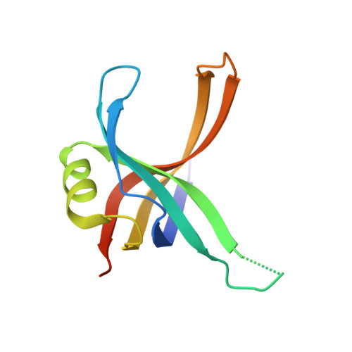
7YM1, 8GW5 - PubMed Abstract:
Single-stranded DNA-binding proteins (SSBs) play a crucial role in DNA metabolism by binding and stabilizing single-stranded DNA (ssDNA) intermediates. Through their multifaceted roles in DNA replication, recombination, repair, replication restart, and other cellular processes, SSB emerges as a central player in maintaining genomic integrity. These attributes collectively position SSBs as essential guardians of genomic integrity, establishing interactions with an array of distinct proteins. Unlike Escherichia coli , which contains only one type of SSB, some bacteria have two paralogous SSBs, referred to as SsbA and SsbB. In this study, we identified Staphylococcus aureus SsbA (SaSsbA) as a fresh addition to the roster of the anticancer drug 5-fluorouracil (5-FU) binding proteins, thereby expanding the ambit of the 5-FU interactome to encompass this DNA replication protein. To investigate the binding mode, we solved the complexed crystal structure with 5-FU at 2.3 Å (PDB ID 7YM1). The structure of glycerol-bound SaSsbA was also determined at 1.8 Å (PDB ID 8GW5). The interaction between 5-FU and SaSsbA was found to involve R18, P21, V52, F54, Q78, R80, E94, and V96. Based on the collective results from mutational and structural analyses, it became evident that SaSsbA's mode of binding with 5-FU diverges from that of SaSsbB. This complexed structure also holds the potential to furnish valuable comprehension regarding how 5-FU might bind to and impede analogous proteins in humans, particularly within cancer-related signaling pathways. Leveraging the information furnished by the glycerol and 5-FU binding sites, the complexed structures of SaSsbA bring to the forefront the potential viability of several interactive residues as potential targets for therapeutic interventions aimed at curtailing SaSsbA activity. Acknowledging the capacity of microbiota to influence the host's response to 5-FU, there emerges a pressing need for further research to revisit the roles that bacterial and human SSBs play in the realm of anticancer therapy.
- Department of Pharmacy, Chia Nan University of Pharmacy and Science, Tainan City 717, Taiwan.
Organizational Affiliation: