Specific binding of Hg 2+ to mismatched base pairs involving 5-hydroxyuracil in duplex DNA.
Torigoe, H., Kondo, J., Arakawa, F.(2023) J Inorg Biochem 241: 112125-112125
- PubMed: 36716510 Search on PubMed
- DOI: https://doi.org/10.1016/j.jinorgbio.2023.112125
- Primary Citation Related Structures:
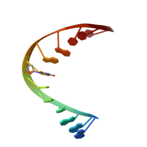
7YGO, 7YGP - PubMed Abstract:
Metal ion-nucleic acid interactions contribute significantly to nucleic acid structure and biological activity and have potential applications in nanotechnology. Hg 2+ specifically binds to the natural T-T mismatched base pair in duplex DNA to form a T-Hg-T base pair. Metal ions may enhance DNA damage induced by DNA-damaging agents, such as oxidative agents. The interactions between metal ions and damaged DNAs, such as mismatched oxidized bases, have not been well characterized. Here, we examined the possibility of Hg 2+ binding to an asymmetric mismatched base pair involving thymine and 5-hydroxyuracil (OHdU), an oxidized base produced by the oxidative deamination of cytosine. UV melting analyses showed that only the melting temperature of the single T-OHdU mismatched duplex DNA increased upon Hg 2+ addition. CD spectra indicated no significant change in the higher-order structure of the single T-OHdU mismatched duplex DNA upon Hg 2+ addition. X-ray crystallographic structure with two consecutive T-OHdU mismatched base pairs and isothermal titration calorimetric analyses with the single T-OHdU mismatched base pair showed that Hg 2+ specifically binds to the N3 positions of both T and OHdU in T-OHdU at 1:1 molar ratio, with a 5×10 5 M -1 binding constant of to form the T-Hg-OHdU base pair. The Hg 2+ -bound structure and the Hg 2+ -binding affinity for T-OHdU was similar to those for T-T. This study on T-Hg-OHdU metal-mediated base pair could aid in studying the molecular mechanism of metal ion-mediated DNA damage and their potential applications in nanotechnology.
- Department of Applied Chemistry, Faculty of Science, Tokyo University of Science, 1-3 Kagurazaka, Shinjuku-ku, Tokyo 162-8601, Japan. Electronic address: htorigoe@rs.tus.ac.jp.
Organizational Affiliation: