7YF1 | pdb_00007yf1

Structure of FABP at 1.7 Angstroms resolution.


Experimental Data Snapshot

  • Method: X-RAY DIFFRACTION
  • Resolution: 1.70 Å
  • R-Value Free: 
    0.197 (Depositor), 0.207 (DCC) 
  • R-Value Work: 
    0.169 (Depositor) 
  • R-Value Observed: 
    0.170 (Depositor) 

Starting Model: experimental
View more details

wwPDB Validation 3D Report Full Report

Validation slider image for 7YF1

Ligand Structure Quality Assessment 


This is version 1.2 of the entry. See complete history

Literature

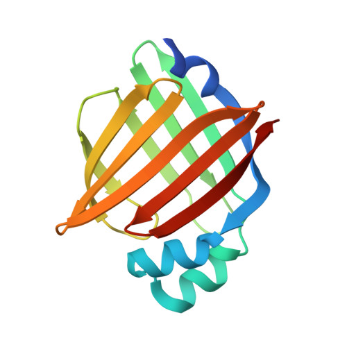
Structural Insights into Mouse H-FABP.

Wang, L.Zhang, H.Lv, P.Li, Y.Teng, M.Liu, Y.Wu, D.

(2022) Life (Basel) 12

  • DOI: https://doi.org/10.3390/life12091445
  • Primary Citation Related Structures: 
    7YF1

  • PubMed Abstract: 

    Intracellular fatty acid-binding proteins are evolutionarily highly conserved proteins. The major functions and responsibilities of this family are the regulation of FA uptake and intracellular transport. The structure of the H-FABP ortholog from mouse ( Mus musculus ) had not been revealed at the time this study was completed. Thus, further exploration of the structural properties of mouse H-FABP is expected to extend our knowledge of the model animal's molecular mechanism of H-FABP function. Here, we report the high-resolution crystal structure and the NMR characterization of mouse H-FABP. Our work discloses the unique structural features of mouse H-FABP, offering a structural basis for the further development of small-molecule inhibitors for H-FABP.


  • Organizational Affiliation
    • School of Life Science, University of Science and Technology of China, Hefei 230027, China.

Macromolecule Content 

  • Total Structure Weight: 16.07 kDa 
  • Atom Count: 1,274 
  • Modeled Residue Count: 133 
  • Deposited Residue Count: 141 
  • Unique protein chains: 1

Macromolecules

Find similar proteins by:|  3D Structure
Entity ID: 1
MoleculeChains  Sequence LengthOrganismDetailsImage
Fatty acid-binding protein, heart141Mus musculusMutation(s): 0 
Gene Names: Fabp3Fabph1
UniProt & NIH Common Fund Data Resources
Find proteins for P11404 (Mus musculus)
Explore P11404 
Go to UniProtKB:  P11404
IMPC:  MGI:95476
Entity Groups
Sequence Clusters30% Identity50% Identity70% Identity90% Identity95% Identity100% Identity
UniProt GroupP11404
Sequence Annotations
Expand
Reference Sequence

Small Molecules

Ligands 1 Unique
IDChains Name / Formula / InChI Key2D Diagram3D Interactions
PLM

Query on PLM


Download:Ideal Coordinates CCD File
B [auth A]PALMITIC ACID
C16 H32 O2
IPCSVZSSVZVIGE-UHFFFAOYSA-N

Experimental Data & Validation

Experimental Data

  • Method: X-RAY DIFFRACTION
  • Resolution: 1.70 Å
  • R-Value Free:  0.197 (Depositor), 0.207 (DCC) 
  • R-Value Work:  0.169 (Depositor) 
  • R-Value Observed: 0.170 (Depositor) 
Space Group: P 21 21 21
Unit Cell:
Length ( Å )Angle ( ˚ )
a = 37.305α = 90
b = 54.419β = 90
c = 65.387γ = 90
Software Package:
Software NamePurpose
PHENIXrefinement
DENZOdata reduction
SCALAdata scaling
PHENIXphasing

Structure Validation

View Full Validation Report


Ligand Structure Quality Assessment 


Entry History 

& Funding Information

Deposition Data


Funding OrganizationLocationGrant Number
Other governmentChinaChina Postdoctoral Science Foundation:2020M682420
Other governmentChinaGuangzhou International Collaborative Grant:2019A050510027

Revision History  (Full details and data files)

  • Version 1.0: 2022-09-21
    Type: Initial release
  • Version 1.1: 2023-04-05
    Changes: Database references
  • Version 1.2: 2023-11-29
    Changes: Data collection, Refinement description