7YE3

Crystal structure of Lactobacillus rhamnosus 4-deoxy-L-threo-5-hexosulose-uronate ketol-isomerase KduI complexed with MES


Experimental Data Snapshot

  • Method: X-RAY DIFFRACTION
  • Resolution: 2.55 Å
  • R-Value Free: 0.256 
  • R-Value Work: 0.193 
  • R-Value Observed: 0.197 

wwPDB Validation   3D Report Full Report


Ligand Structure Quality Assessment 


This is version 1.3 of the entry. See complete history


Literature

Crystal Structures of Lacticaseibacillus 4-Deoxy-L- threo- 5-hexosulose-uronate Ketol-isomerase KduI in Complex with Substrate Analogs.

Iwase, H.Yamamoto, Y.Yamada, A.Kawai, K.Oiki, S.Watanabe, D.Mikami, B.Takase, R.Hashimoto, W.

(2023) J Appl Glycosci (1999) 70: 99-107

  • DOI: https://doi.org/10.5458/jag.jag.JAG-2023_0003
  • Primary Citation of Related Structures:  
    7YE3, 7YRS

  • PubMed Abstract: 

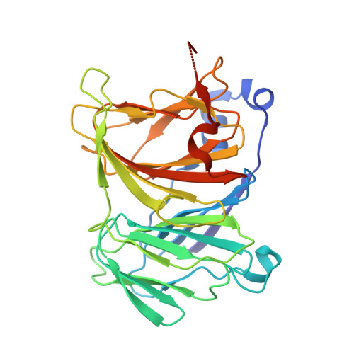
    Some probiotics including lactobacilli, colonize host animal cells by targeting glycosaminoglycans (GAGs), such as heparin, located in the extracellular matrix. Recent studies have shown that several lactic acid bacteria degrade GAGs. Here we show the structure/function relationship of Lacticaseibacillus rhamnosus 4-deoxy-L- threo -5-hexosulose-uronate ketol-isomerase (KduI) crucial for metabolism of unsaturated glucuronic acid produced through degradation of GAGs. Crystal structures of ligand-free and bound KduIs were determined by X-ray crystallography and the enzyme was found to consist of six identical subunits and adopt a β-helix as a basic scaffold. Ligands structurally similar to the substrate were bound to the cleft of each enzyme subunit. Several residues located in the cleft interacted with ligands through hydrogen bonds and/or C-C contacts. In addition to substrate analogs, a metal ion coordinated to four residues, His198, His200, Glu205, and His248, in the cleft, and the enzyme activity was significantly inhibited by a chelator, ethylenediaminetetraacetic acid. Site-directed mutants in Arg163, Ile165, Thr184, Thr194, His200, Arg203, Tyr207, Met262, and Tyr269 in the cleft exhibited little enzyme activity, indicating that these residues and the metal ion constituted an active site in the cleft. This is the first report on the active site structure of KduI based on the ligand-bound complex.


  • Organizational Affiliation

    1 Laboratory of Basic and Applied Molecular Biotechnology, Division of Food Science and Biotechnology, Graduate School of Agriculture, Kyoto University.


Macromolecules
Find similar proteins by:  (by identity cutoff)  |  3D Structure
Entity ID: 1
MoleculeChains Sequence LengthOrganismDetailsImage
4-deoxy-L-threo-5-hexosulose-uronate ketol-isomerase
A, B, C, D, E
A, B, C, D, E, F
289Lacticaseibacillus rhamnosusMutation(s): 0 
Gene Names: kduIHMPREF0539_0625
EC: 5.3.1.17
UniProt
Find proteins for C2JUP1 (Lacticaseibacillus rhamnosus (strain LMS2-1))
Explore C2JUP1 
Go to UniProtKB:  C2JUP1
Entity Groups  
Sequence Clusters30% Identity50% Identity70% Identity90% Identity95% Identity100% Identity
UniProt GroupC2JUP1
Sequence Annotations
Expand
  • Reference Sequence
Small Molecules
Ligands 2 Unique
IDChains Name / Formula / InChI Key2D Diagram3D Interactions
MES (Subject of Investigation/LOI)
Query on MES

Download Ideal Coordinates CCD File 
H [auth A],
J [auth B],
L [auth C],
P [auth F]
2-(N-MORPHOLINO)-ETHANESULFONIC ACID
C6 H13 N O4 S
SXGZJKUKBWWHRA-UHFFFAOYSA-N
ZN (Subject of Investigation/LOI)
Query on ZN

Download Ideal Coordinates CCD File 
G [auth A]
I [auth B]
K [auth C]
M [auth D]
N [auth E]
G [auth A],
I [auth B],
K [auth C],
M [auth D],
N [auth E],
O [auth F]
ZINC ION
Zn
PTFCDOFLOPIGGS-UHFFFAOYSA-N
Experimental Data & Validation

Experimental Data

  • Method: X-RAY DIFFRACTION
  • Resolution: 2.55 Å
  • R-Value Free: 0.256 
  • R-Value Work: 0.193 
  • R-Value Observed: 0.197 
  • Space Group: C 1 2 1
Unit Cell:
Length ( Å )Angle ( ˚ )
a = 93.035α = 90
b = 189.102β = 107.12
c = 114.852γ = 90
Software Package:
Software NamePurpose
PHENIXrefinement
PDB_EXTRACTdata extraction
XDSdata reduction
Aimlessdata scaling
MOLREPphasing

Structure Validation

View Full Validation Report


Ligand Structure Quality Assessment 


Entry History & Funding Information

Deposition Data


Funding OrganizationLocationGrant Number
Japan Society for the Promotion of Science (JSPS)Japan--

Revision History  (Full details and data files)

  • Version 1.0: 2023-07-05
    Type: Initial release
  • Version 1.1: 2023-11-15
    Changes: Data collection, Database references
  • Version 1.2: 2023-11-29
    Changes: Refinement description
  • Version 1.3: 2024-01-31
    Changes: Database references