7YA7 | pdb_00007ya7

The crystal structure of IpaH1.4 LRR domain


Experimental Data Snapshot

  • Method: X-RAY DIFFRACTION
  • Resolution: 1.40 Å
  • R-Value Free: 
    0.220 (Depositor), 0.221 (DCC) 
  • R-Value Work: 
    0.185 (Depositor), 0.186 (DCC) 
  • R-Value Observed: 
    0.187 (Depositor) 

Starting Model: experimental
View more details

wwPDB Validation 3D Report Full Report

Validation slider image for 7YA7

This is version 1.3 of the entry. See complete history

Literature

Structural insight into the recognition of the linear ubiquitin assembly complex by Shigella E3 ligase IpaH1.4/2.5.

Hiragi, K.Nishide, A.Takagi, K.Iwai, K.Kim, M.Mizushima, T.

(2023) J Biochem 173: 317-326

  • DOI: https://doi.org/10.1093/jb/mvac109
  • Primary Citation Related Structures: 
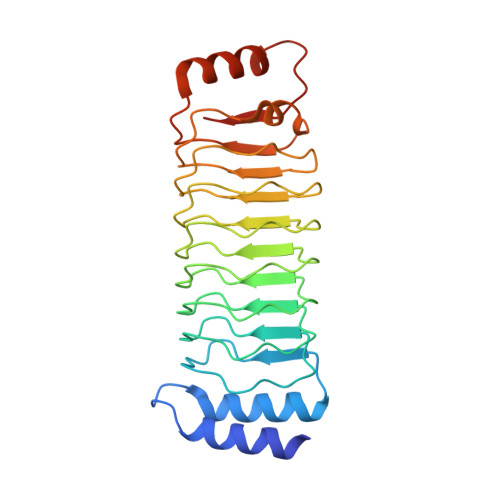
    7YA7, 7YA8

  • PubMed Abstract: 

    Pathogenic bacteria deliver virulence factors called effectors into host cells in order to facilitate infection. The Shigella effector proteins IpaH1.4 and IpaH2.5 are members of the 'novel E3 ligase' (NEL)-type bacterial E3 ligase family. These proteins ubiquitinate the linear ubiquitin assembly complex (LUBAC) to inhibit nuclear factor (NF)-κB activation and, concomitantly, the inflammatory response. However, the molecular mechanisms underlying the interaction and recognition between IpaH1.4 and IpaH2.5 and LUBAC are unclear. Here we present the crystal structures of the substrate-recognition domains of IpaH1.4 and IpaH2.5 at resolutions of 1.4 and 3.4 Å, respectively. The LUBAC-binding site on IpaH1.4 was predicted based on structural comparisons with the structures of other NEL-type E3s. Structural and biochemical data were collected and analysed to determine the specific residues of IpaH1.4 that are involved in interactions with LUBAC and influence NF-κB signaling. The new structural insight presented here demonstrates how bacterial pathogens target innate immune signaling pathways.


  • Organizational Affiliation
    • Department of Science, Graduate School of Science, University of Hyogo, 2167, Shosha, Himeji, Hyogo, 671-2280, Japan.

Macromolecule Content 

  • Total Structure Weight: 54.64 kDa 
  • Atom Count: 4,150 
  • Modeled Residue Count: 454 
  • Deposited Residue Count: 486 
  • Unique protein chains: 1

Macromolecules

Find similar proteins by:|  3D Structure
Entity ID: 1
MoleculeChains  Sequence LengthOrganismDetailsImage
RING-type E3 ubiquitin transferase
A, B
243Shigella flexneri 5a str. M90TMutation(s): 0 
Gene Names: ipaH1.4S0283pWR501_0283
EC: 2.3.2.27
UniProt
Find proteins for A0A0H2USG1 (Shigella flexneri)
Explore A0A0H2USG1 
Go to UniProtKB:  A0A0H2USG1
Entity Groups
Sequence Clusters30% Identity50% Identity70% Identity90% Identity95% Identity100% Identity
UniProt GroupA0A0H2USG1
Sequence Annotations
Expand
Reference Sequence

Experimental Data & Validation

Experimental Data

  • Method: X-RAY DIFFRACTION
  • Resolution: 1.40 Å
  • R-Value Free:  0.220 (Depositor), 0.221 (DCC) 
  • R-Value Work:  0.185 (Depositor), 0.186 (DCC) 
  • R-Value Observed: 0.187 (Depositor) 
Space Group: P 1 21 1
Unit Cell:
Length ( Å )Angle ( ˚ )
a = 33.467α = 90
b = 68.039β = 100.72
c = 100.26γ = 90
Software Package:
Software NamePurpose
REFMACrefinement
HKL-2000data collection
HKL-2000data scaling
MOLREPphasing
HKL-2000data reduction

Structure Validation

View Full Validation Report


Entry History 

& Funding Information

Deposition Data


Funding OrganizationLocationGrant Number
Japan Society for the Promotion of Science (JSPS)JapanJP24112009

Revision History  (Full details and data files)

  • Version 1.0: 2023-02-08
    Type: Initial release
  • Version 1.1: 2023-02-22
    Changes: Refinement description
  • Version 1.2: 2023-04-12
    Changes: Database references
  • Version 1.3: 2023-11-29
    Changes: Data collection, Refinement description