7Y8Q | pdb_00007y8q

Amyloid-beta assemblage on GM1-containing membranes


Experimental Data Snapshot

  • Method: SOLID-STATE NMR
  • Conformers Calculated: 10 
  • Conformers Submitted: 10 
  • Selection Criteria: structures with the lowest energy 

wwPDB Validation 3D Report Full Report

Validation slider image for 7Y8Q

This is version 1.2 of the entry. See complete history

Literature

The Double-Layered Structure of Amyloid-beta Assemblage on GM1-Containing Membranes Catalytically Promotes Fibrillization.

Yagi-Utsumi, M.Itoh, S.G.Okumura, H.Yanagisawa, K.Kato, K.Nishimura, K.

(2023) ACS Chem Neurosci 14: 2648-2657

  • DOI: https://doi.org/10.1021/acschemneuro.3c00192
  • Primary Citation Related Structures: 
    7Y8Q

  • PubMed Abstract: 

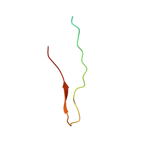
    Alzheimer's disease (AD) is associated with progressive accumulation of amyloid-β (Aβ) cross-β fibrils in the brain. Aβ species tightly associated with GM1 ganglioside, a glycosphingolipid abundant in neuronal membranes, promote amyloid fibril formation; therefore, they could be attractive clinical targets. However, the active conformational state of Aβ in GM1-containing lipid membranes is still unknown. The present solid-state nuclear magnetic resonance study revealed a nonfibrillar Aβ assemblage characterized by a double-layered antiparallel β-structure specifically formed on GM1 ganglioside clusters. Our data show that this unique assemblage was not transformed into fibrils on GM1-containing membranes but could promote conversion of monomeric Aβ into fibrils, suggesting that a solvent-exposed hydrophobic layer provides a catalytic surface evoking Aβ fibril formation. Our findings offer structural clues for designing drugs targeting catalytically active Aβ conformational species for the development of anti-AD therapeutics.


  • Organizational Affiliation
    • Exploratory Research Center on Life and Living Systems (ExCELLS), National Institutes of Natural Sciences, Okazaki, Aichi 444-8787, Japan.

Macromolecule Content 

  • Total Structure Weight: 34.69 kDa 
  • Atom Count: 1,704 
  • Modeled Residue Count: 232 
  • Deposited Residue Count: 320 
  • Unique protein chains: 1

Macromolecules

Find similar proteins by:|  3D Structure
Entity ID: 1
MoleculeChains  Sequence LengthOrganismDetailsImage
Amyloid-beta protein 40
A, B, C, D, E
A, B, C, D, E, F, G, H
40Homo sapiensMutation(s): 0 
Gene Names: APPA4AD1
UniProt & NIH Common Fund Data Resources
Find proteins for P05067 (Homo sapiens)
Explore P05067 
Go to UniProtKB:  P05067
PHAROS:  P05067
GTEx:  ENSG00000142192 
Entity Groups
Sequence Clusters30% Identity50% Identity70% Identity90% Identity95% Identity100% Identity
UniProt GroupP05067
Sequence Annotations
Expand
Reference Sequence

Experimental Data & Validation

Experimental Data

  • Method: SOLID-STATE NMR
  • Conformers Calculated: 10 
  • Conformers Submitted: 10 
  • Selection Criteria: structures with the lowest energy 

Structure Validation

View Full Validation Report


Entry History 

& Funding Information

Deposition Data


Funding OrganizationLocationGrant Number
Japan Society for the Promotion of Science (JSPS)JapanJP19K07041
Japan Society for the Promotion of Science (JSPS)JapanJP19K05552

Revision History  (Full details and data files)

  • Version 1.0: 2023-07-05
    Type: Initial release
  • Version 1.1: 2023-09-20
    Changes: Data collection, Database references
  • Version 1.2: 2024-05-15
    Changes: Database references