7Y68 | pdb_00007y68

SIDT2-pH5.5 plus miRNA


Experimental Data Snapshot

  • Method: ELECTRON MICROSCOPY
  • Resolution: 2.87 Å
  • Aggregation State: PARTICLE 
  • Reconstruction Method: SINGLE PARTICLE 

wwPDB Validation 3D Report Full Report

Validation slider image for 7Y68

This is version 1.2 of the entry. See complete history

Literature

Structural insight into the human SID1 transmembrane family member 2 reveals its lipid hydrolytic activity.

Qian, D.Cong, Y.Wang, R.Chen, Q.Yan, C.Gong, D.S.

(2023) Nat Commun 14: 3568-3568

  • DOI: https://doi.org/10.1038/s41467-023-39335-2
  • Primary Citation Related Structures: 
    7Y63, 7Y68, 7Y69

  • PubMed Abstract: 

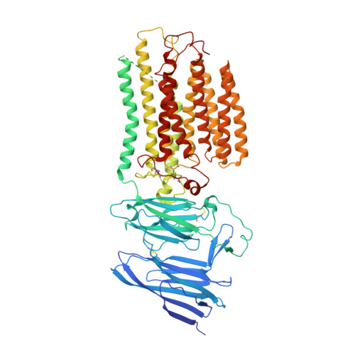
    The systemic RNAi-defective (SID) transmembrane family member 2 (SIDT2) is a putative nucleic acid channel or transporter that plays essential roles in nucleic acid transport and lipid metabolism. Here, we report the cryo-electron microscopy (EM) structures of human SIDT2, which forms a tightly packed dimer with extensive interactions mediated by two previously uncharacterized extracellular/luminal β-strand-rich domains and the unique transmembrane domain (TMD). The TMD of each SIDT2 protomer contains eleven transmembrane helices (TMs), and no discernible nucleic acid conduction pathway has been identified within the TMD, suggesting that it may act as a transporter. Intriguingly, TM3-6 and TM9-11 form a large cavity with a putative catalytic zinc atom coordinated by three conserved histidine residues and one aspartate residue lying approximately 6 Å from the extracellular/luminal surface of the membrane. Notably, SIDT2 can hydrolyze C18 ceramide into sphingosine and fatty acid with a slow rate. The information presented advances the understanding of the structure-function relationships in the SID1 family proteins.


  • Organizational Affiliation
    • State Key Laboratory of Medicinal Chemical Biology and College of Life Sciences, Nankai University, Tianjin, 300350, China.

Macromolecule Content 

  • Total Structure Weight: 192.68 kDa 
  • Atom Count: 10,264 
  • Modeled Residue Count: 1,300 
  • Deposited Residue Count: 1,664 
  • Unique protein chains: 1

Macromolecules

Find similar proteins by:|  3D Structure
Entity ID: 1
MoleculeChains  Sequence LengthOrganismDetailsImage
SID1 transmembrane family member 2
A, B
832Homo sapiensMutation(s): 0 
Gene Names: SIDT2CGI-40PSEC0072UNQ685/PRO1325
Membrane Entity: Yes 
UniProt & NIH Common Fund Data Resources
Find proteins for Q8NBJ9 (Homo sapiens)
Explore Q8NBJ9 
Go to UniProtKB:  Q8NBJ9
PHAROS:  Q8NBJ9
GTEx:  ENSG00000149577 
Entity Groups
Sequence Clusters30% Identity50% Identity70% Identity90% Identity95% Identity100% Identity
UniProt GroupQ8NBJ9
Glycosylation
Glycosylation Sites: 6Go to GlyGen: Q8NBJ9-1
Sequence Annotations
Expand
Reference Sequence

Oligosaccharides

Help  
Entity ID: 2
MoleculeChains Length2D Diagram GlycosylationD Interactions
2-acetamido-2-deoxy-beta-D-glucopyranose-(1-4)-2-acetamido-2-deoxy-beta-D-glucopyranose
C, D, E, F
2N-Glycosylation
Glycosylation Resources
GlyTouCan: G42666HT
GlyCosmos: G42666HT
GlyGen: G42666HT

Small Molecules

Ligands 2 Unique
IDChains Name / Formula / InChI Key2D Diagram3D Interactions
NAG
(Subject of Investigation/LOI)

Query on NAG


Download:Ideal Coordinates CCD File
H [auth A]
I [auth A]
J [auth A]
K [auth A]
M [auth B]
H [auth A],
I [auth A],
J [auth A],
K [auth A],
M [auth B],
N [auth B],
O [auth B],
P [auth B]
2-acetamido-2-deoxy-beta-D-glucopyranose
C8 H15 N O6
OVRNDRQMDRJTHS-FMDGEEDCSA-N
ZN
(Subject of Investigation/LOI)

Query on ZN


Download:Ideal Coordinates CCD File
G [auth A],
L [auth B]
ZINC ION
Zn
PTFCDOFLOPIGGS-UHFFFAOYSA-N

Experimental Data & Validation

Experimental Data

  • Method: ELECTRON MICROSCOPY
  • Resolution: 2.87 Å
  • Aggregation State: PARTICLE 
  • Reconstruction Method: SINGLE PARTICLE 

Structure Validation

View Full Validation Report


Entry History 

& Funding Information

Deposition Data

  • Released Date: 2023-06-21 
  • Deposition Author(s): Gong, D.S.

Funding OrganizationLocationGrant Number
Not funded--

Revision History  (Full details and data files)

  • Version 1.0: 2023-06-21
    Type: Initial release
  • Version 1.1: 2023-11-29
    Changes: Data collection, Database references
  • Version 1.2: 2024-11-06
    Changes: Data collection, Structure summary