Hybrid in vitro/in silico analysis of low-affinity protein-protein interactions that regulate signal transduction by Sema6D.
Tanaka, T., Ekimoto, T., Nagatomo, M., Neyazaki, M., Shimoji, E., Yamane, T., Kanagawa, S., Oi, R., Mihara, E., Takagi, J., Akashi, S., Ikeguchi, M., Nogi, T.(2022) Protein Sci 31: e4452-e4452
- PubMed: 36156831 Search on PubMedSearch on PubMed Central
- DOI: https://doi.org/10.1002/pro.4452
- Primary Citation Related Structures:
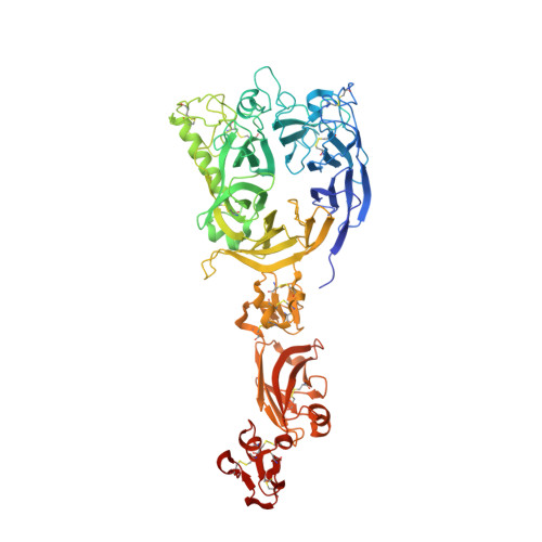
7Y4O, 7Y4P, 7Y4Q - PubMed Abstract:
Semaphorins constitute a large family of secreted and membrane-bound proteins that signal through cell-surface receptors, plexins. Semaphorins generally use low-affinity protein-protein interactions to bind with their specific plexin(s) and regulate distinct cellular processes such as neurogenesis, immune response, and organogenesis. Sema6D is a membrane-bound semaphorin that interacts with class A plexins. Sema6D exhibited differential binding affinities to class A plexins in prior cell-based assays, but the molecular mechanism underlying this selectivity is not well understood. Therefore, we performed hybrid in vitro/in silico analysis to examine the binding mode of Sema6D to class A plexins and to identify residues that give rise to the differential affinities and thus contribute to the selectivity within the same class of semaphorins. Our biophysical binding analysis indeed confirmed that Sema6D has a higher affinity for Plexin-A1 than for other class A plexins, consistent with the binding selectivity observed in the previous cell-based assays. Unexpectedly, our present crystallographic analysis of the Sema6D-Plexin-A1 complex showed that the pattern of polar interactions is not interaction-specific because it matches the pattern in the prior structure of the Sema6A-Plexin-A2 complex. Thus, we performed in silico alanine scanning analysis and discovered hotspot residues that selectively stabilized the Sema6D-Plexin-A1 pair via Van der Waals interactions. We then validated the contribution of these hotspot residues to the variation in binding affinity with biophysical binding analysis and molecular dynamics simulations on the mutants. Ultimately, our present results suggest that shape complementarity in the binding interfaces is a determinant for binding selectivity.
- Graduate School of Medical Life Science, Yokohama City University, Yokohama, Kanagawa, Japan.
Organizational Affiliation: