7Y4M

Crystal structure of Ricin A chain bound with N2-(2-amino-4-oxo-3,4-dihydropteridine-7-carbonyl)glycyl-L-phenylalanyl-N6-((benzyloxy)carbonyl)-L-lysine


Experimental Data Snapshot

  • Method: X-RAY DIFFRACTION
  • Resolution: 1.45 Å
  • R-Value Free: 0.242 
  • R-Value Work: 0.222 
  • R-Value Observed: 0.223 

wwPDB Validation   3D Report Full Report


Ligand Structure Quality Assessment 


This is version 1.2 of the entry. See complete history


Literature

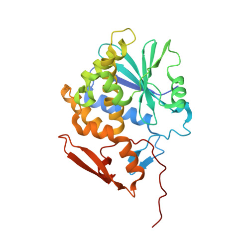
Pterin-based small molecule inhibitor capable of binding to the secondary pocket in the active site of ricin-toxin A chain.

Saito, R.Goto, M.Katakura, S.Ohba, T.Kawata, R.Nagatsu, K.Higashi, S.Kurisu, K.Matsumoto, K.Ohtsuka, K.

(2022) PLoS One 17: e0277770-e0277770

  • DOI: https://doi.org/10.1371/journal.pone.0277770
  • Primary Citation of Related Structures:  
    7Y4K, 7Y4M

  • PubMed Abstract: 

    The Ricin toxin A chain (RTA), which depurinates an adenine base at a specific region of the ribosome leading to death, has two adjacent specificity pockets in its active site. Based on this structural information, many attempts have been made to develop small-molecule RTA inhibitors that simultaneously block the two pockets. However, no attempt has been successful. In the present study, we synthesized pterin-7-carboxamides with tripeptide pendants and found that one of them interacts with both pockets simultaneously to exhibit good RTA inhibitory activity. X-ray crystallographic analysis of the RTA crystal with the new inhibitor revealed that the conformational change of Tyr80 is an important factor that allows the inhibitors to plug the two pockets simultaneously.


  • Organizational Affiliation

    Department of Chemistry, Faculty of Science, Toho University, Funabashi, Chiba, Japan.


Macromolecules
Find similar proteins by:  (by identity cutoff)  |  3D Structure
Entity ID: 1
MoleculeChains Sequence LengthOrganismDetailsImage
Ricin A chain274Ricinus communisMutation(s): 0 
EC: 3.2.2.22
UniProt
Find proteins for P02879 (Ricinus communis)
Explore P02879 
Go to UniProtKB:  P02879
Entity Groups  
Sequence Clusters30% Identity50% Identity70% Identity90% Identity95% Identity100% Identity
UniProt GroupP02879
Sequence Annotations
Expand
  • Reference Sequence
Small Molecules
Ligands 2 Unique
IDChains Name / Formula / InChI Key2D Diagram3D Interactions
IWQ (Subject of Investigation/LOI)
Query on IWQ

Download Ideal Coordinates CCD File 
B [auth A](2S)-2-[[(2S)-2-[2-[(2-azanyl-4-oxidanylidene-3H-pteridin-7-yl)carbonylamino]ethanoylamino]-3-phenyl-propanoyl]amino]-6-(phenylmethoxycarbonylamino)hexanoic acid
C32 H35 N9 O8
FVYSJCKPWXGJBU-VXKWHMMOSA-N
SO4
Query on SO4

Download Ideal Coordinates CCD File 
C [auth A],
D [auth A]
SULFATE ION
O4 S
QAOWNCQODCNURD-UHFFFAOYSA-L
Experimental Data & Validation

Experimental Data

  • Method: X-RAY DIFFRACTION
  • Resolution: 1.45 Å
  • R-Value Free: 0.242 
  • R-Value Work: 0.222 
  • R-Value Observed: 0.223 
  • Space Group: P 41 21 2
Unit Cell:
Length ( Å )Angle ( ˚ )
a = 67.752α = 90
b = 67.752β = 90
c = 141.316γ = 90
Software Package:
Software NamePurpose
XDSdata reduction
SCALAdata scaling
MOLREPphasing
REFMACrefinement
PDB_EXTRACTdata extraction

Structure Validation

View Full Validation Report


Ligand Structure Quality Assessment 


Entry History & Funding Information

Deposition Data


Funding OrganizationLocationGrant Number
Japan Society for the Promotion of Science (JSPS)JapanJP19K05699

Revision History  (Full details and data files)

  • Version 1.0: 2022-11-16
    Type: Initial release
  • Version 1.1: 2022-12-21
    Changes: Database references
  • Version 1.2: 2023-11-29
    Changes: Data collection, Refinement description