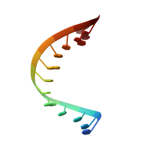
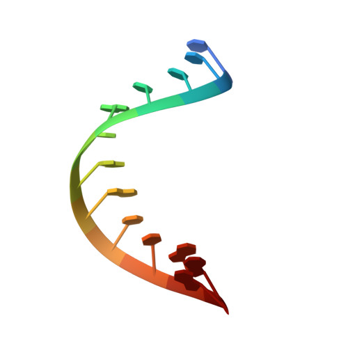
Structural basis for water modulating RNA duplex formation in the CUG repeats of myotonic dystrophy type 1.
Wang, S.C., Chen, Y.T., Satange, R., Chu, J.W., Hou, M.H.(2023) J Biological Chem 299: 104864-104864
- PubMed: 37245780 Search on PubMedSearch on PubMed Central
- DOI: https://doi.org/10.1016/j.jbc.2023.104864
- Primary Citation Related Structures:
7Y2B, 7Y2P - PubMed Abstract:
Secondary structures formed by expanded CUG RNA are involved in the pathobiology of myotonic dystrophy type 1. Understanding the molecular basis of toxic RNA structures can provide insights into the mechanism of disease pathogenesis and accelerate the drug discovery process. Here, we report the crystal structure of CUG repeat RNA containing three U-U mismatches between C-G and G-C base pairs. The CUG RNA crystallizes as an A-form duplex, with the first and third U-U mismatches adopting a water-mediated asymmetric mirror isoform geometry. We found for the first time that a symmetric, water-bridged U-H 2 O-U mismatch is well tolerated within the CUG RNA duplex, which was previously suspected but not observed. The new water-bridged U-U mismatch resulted in high base-pair opening and single-sided cross-strand stacking interactions, which in turn dominate the CUG RNA structure. Furthermore, we performed molecular dynamics simulations that complemented the structural findings and proposed that the first and third U-U mismatches are interchangeable conformations, while the central water-bridged U-U mismatch represents an intermediate state that modulates the RNA duplex conformation. Collectively, the new structural features provided in this work are important for understanding the recognition of U-U mismatches in CUG repeats by external ligands such as proteins or small molecules.
- Institute of Genomics and Bioinformatics, National Chung Hsing University, Taichung, Taiwan; PhD. Program in Medical Biotechnology, National Chung Hsing University, Taichung, Taiwan.
Organizational Affiliation: