7Y13 | pdb_00007y13

Cryo-EM structure of apo-state MrgD-Gi complex (local)


Experimental Data Snapshot

  • Method: ELECTRON MICROSCOPY
  • Resolution: 3.10 Å
  • Aggregation State: PARTICLE 
  • Reconstruction Method: SINGLE PARTICLE 

wwPDB Validation 3D Report Full Report

Validation slider image for 7Y13

This is version 1.2 of the entry. See complete history

Literature

Structural insight into the activation mechanism of MrgD with heterotrimeric Gi-protein revealed by cryo-EM.

Suzuki, S.Iida, M.Hiroaki, Y.Tanaka, K.Kawamoto, A.Kato, T.Oshima, A.

(2022) Commun Biol 5: 707-707

  • DOI: https://doi.org/10.1038/s42003-022-03668-3
  • Primary Citation Related Structures: 
    7Y12, 7Y13, 7Y14, 7Y15

  • PubMed Abstract: 

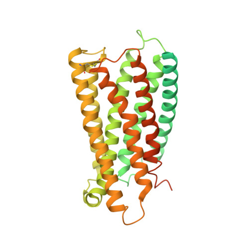
    MrgD, a member of the Mas-related G protein-coupled receptor (MRGPR) family, has high basal activity for Gi activation. It recognizes endogenous ligands, such as β-alanine, and is involved in pain and itch signaling. The lack of a high-resolution structure for MrgD hinders our understanding of whether its activation is ligand-dependent or constitutive. Here, we report two cryo-EM structures of the MrgD-Gi complex in the β-alanine-bound and apo states at 3.1 Å and 2.8 Å resolution, respectively. These structures show that β-alanine is bound to a shallow pocket at the extracellular domains. The extracellular half of the sixth transmembrane helix undergoes a significant movement and is tightly packed into the third transmembrane helix through hydrophobic residues, creating the active form. Our structures demonstrate a structural basis for the characteristic ligand recognition of MrgD. These findings provide a framework to guide drug designs targeting the MrgD receptor.


  • Organizational Affiliation
    • Department of Basic Medicinal Sciences, Graduate School of Pharmaceutical Sciences, Nagoya University, Furo-cho, Chikusa-ku, Nagoya, 464-8601, Japan.

Macromolecule Content 

  • Total Structure Weight: 51.94 kDa 
  • Atom Count: 2,199 
  • Modeled Residue Count: 264 
  • Deposited Residue Count: 446 
  • Unique protein chains: 1

Macromolecules

Find similar proteins by:|  3D Structure
Entity ID: 1
MoleculeChains  Sequence LengthOrganismDetailsImage
Soluble cytochrome b562,Mas-related G-protein coupled receptor member DA [auth R]446Escherichia coliHomo sapiens
This entity is chimeric
Mutation(s): 2 
Gene Names: cybCMRGPRDMRGD
Membrane Entity: Yes 
UniProt & NIH Common Fund Data Resources
Find proteins for P0ABE7 (Escherichia coli)
Explore P0ABE7 
Go to UniProtKB:  P0ABE7
Find proteins for Q8TDS7 (Homo sapiens)
Explore Q8TDS7 
Go to UniProtKB:  Q8TDS7
GTEx:  ENSG00000172938 
Entity Groups
Sequence Clusters30% Identity50% Identity70% Identity90% Identity95% Identity100% Identity
UniProt GroupsP0ABE7Q8TDS7
Sequence Annotations
Expand
Reference Sequence

Experimental Data & Validation

Experimental Data

  • Method: ELECTRON MICROSCOPY
  • Resolution: 3.10 Å
  • Aggregation State: PARTICLE 
  • Reconstruction Method: SINGLE PARTICLE 
EM Software:
TaskSoftware PackageVersion
MODEL REFINEMENTPHENIX

Structure Validation

View Full Validation Report


Entry History 

& Funding Information

Deposition Data


Funding OrganizationLocationGrant Number
Japan Society for the Promotion of Science (JSPS)JapanJP19H03165
Japan Agency for Medical Research and Development (AMED)JapanJP21am0101074

Revision History  (Full details and data files)

  • Version 1.0: 2022-07-20
    Type: Initial release
  • Version 1.1: 2022-11-23
    Changes: Database references
  • Version 1.2: 2024-10-30
    Changes: Data collection, Structure summary