From Hit to Lead: Structure-Based Optimization of Novel Selective Inhibitors of Receptor-Interacting Protein Kinase 1 (RIPK1) for the Treatment of Inflammatory Diseases.
Zhang, L., Li, Y., Tian, C., Yang, R., Wang, Y., Xu, H., Zhu, Q., Chen, S., Li, L., Yang, S.(2024) J Med Chem 67: 754-773
- PubMed: 38159286 Search on PubMed
- DOI: https://doi.org/10.1021/acs.jmedchem.3c02102
- Primary Citation Related Structures:
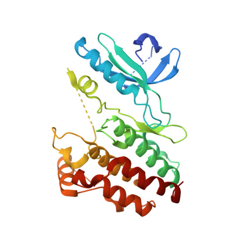
7XMK - PubMed Abstract:
Receptor-interacting protein kinase 1 (RIPK1) is a key regulator of cellular necroptosis, which is considered as an important therapeutic target for necroptosis-related indications. Herein, we report the structural optimization and structure-activity relationship investigations of a series of eutectic 5-substituted-indole-3-carboxamide derivatives. The prioritized compound 10b exhibited low nanomolar IC 50 values against RIPK1 and showed good kinase selectivity. Based on its eutectic structure, 10b occupied both the allosteric and ATP binding pockets of RIPK1, making it a potent dual-mode inhibitor of RIPK1. In vitro , 10b had a potent protective effect against necroptosis in cells. Compound 10b also provided robust protection in a TNFα-induced systemic inflammatory response syndrome (SIRS) model and imiquimod (IMQ)-induced psoriasis model. It also showed good pharmacokinetic properties and low toxicity. Overall, 10b is a promising lead compound for drug discovery targeting RIPK1 and warrants further study.
- Department of Biotherapy, Cancer Center and State Key Laboratory of Biotherapy, West China Hospital, Sichuan University, Chengdu 610041, China.
Organizational Affiliation: