


Discovery of a novel azaindole derivatives targeting the influenza PB2 cap binding region
Zhang, Z., Liu, X.To be published.
Experimental Data Snapshot
Starting Model: experimental
View more details
Entity ID: 1 | |||||
|---|---|---|---|---|---|
| Molecule | Chains | Sequence Length | Organism | Details | Image |
| Polymerase basic protein 2 | 166 | Influenza A virus (A/Victoria/3/1975(H3N2)) | Mutation(s): 0 Gene Names: PB2 | 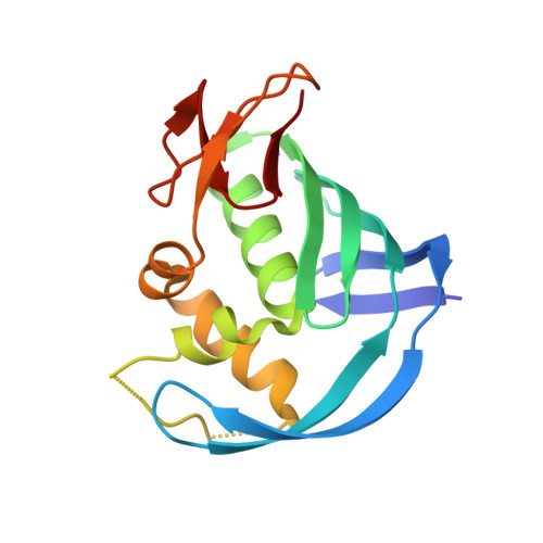 | |
UniProt | |||||
Entity Groups | |||||
| Sequence Clusters | 30% Identity50% Identity70% Identity90% Identity95% Identity100% Identity | ||||
| UniProt Group | P31345 | ||||
Sequence AnnotationsExpand | |||||
Reference Sequence | |||||
| Ligands 2 Unique | |||||
|---|---|---|---|---|---|
| ID | Chains | Name / Formula / InChI Key | 2D Diagram | 3D Interactions | |
| GIH (Subject of Investigation/LOI) Download:Ideal Coordinates CCD File | F [auth A], H [auth B], J [auth C], L [auth D] | (2~{S},3~{S})-3-[[5-dimethoxyphosphoryl-4-(5-fluoranyl-1~{H}-pyrrolo[2,3-b]pyridin-3-yl)pyrimidin-2-yl]amino]bicyclo[2.2.2]octane-2-carboxylic acid C22 H25 F N5 O5 P HZZFRSXJRJLLCP-CMKMFDCUSA-N |  | ||
| IOD Download:Ideal Coordinates CCD File | E [auth A], G [auth B], I [auth C], K [auth D] | IODIDE ION I XMBWDFGMSWQBCA-UHFFFAOYSA-M |  | ||
| Length ( Å ) | Angle ( ˚ ) |
|---|---|
| a = 176.791 | α = 90 |
| b = 31.306 | β = 95.31 |
| c = 114.552 | γ = 90 |
| Software Name | Purpose |
|---|---|
| PHENIX | refinement |
| PDB_EXTRACT | data extraction |
| XDS | data reduction |
| Aimless | data scaling |
| PHASER | phasing |
| Funding Organization | Location | Grant Number |
|---|---|---|
| Not funded | -- |


