7XLQ | pdb_00007xlq

Structure of human R-type voltage-gated CaV2.3-alpha2/delta1-beta1 channel complex in the ligand-free (apo) state


Experimental Data Snapshot

  • Method: ELECTRON MICROSCOPY
  • Resolution: 3.10 Å
  • Aggregation State: PARTICLE 
  • Reconstruction Method: SINGLE PARTICLE 

Starting Model: experimental
View more details

wwPDB Validation 3D Report Full Report

Validation slider image for 7XLQ

This is version 1.1 of the entry. See complete history

Literature

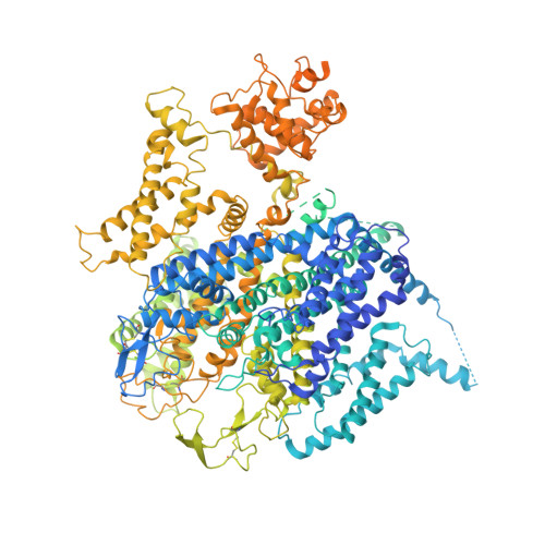
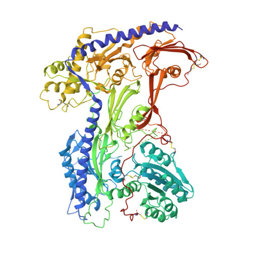
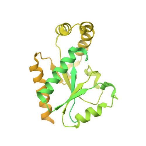
Molecular insights into the gating mechanisms of voltage-gated calcium channel Ca V 2.3.

Gao, Y.Xu, S.Cui, X.Xu, H.Qiu, Y.Wei, Y.Dong, Y.Zhu, B.Peng, C.Liu, S.Zhang, X.C.Sun, J.Huang, Z.Zhao, Y.

(2023) Nat Commun 14: 516-516

  • DOI: https://doi.org/10.1038/s41467-023-36260-2
  • Primary Citation Related Structures: 
    7XLQ

  • PubMed Abstract: 

    High-voltage-activated R-type Ca V 2.3 channel plays pivotal roles in many physiological activities and is implicated in epilepsy, convulsions, and other neurodevelopmental impairments. Here, we determine the high-resolution cryo-electron microscopy (cryo-EM) structure of human Ca V 2.3 in complex with the α2δ1 and β1 subunits. The VSD II is stabilized in the resting state. Electrophysiological experiments elucidate that the VSD II is not required for channel activation, whereas the other VSDs are essential for channel opening. The intracellular gate is blocked by the W-helix. A pre-W-helix adjacent to the W-helix can significantly regulate closed-state inactivation (CSI) by modulating the association and dissociation of the W-helix with the gate. Electrostatic interactions formed between the negatively charged domain on S6 II , which is exclusively conserved in the Ca V 2 family, and nearby regions at the alpha-interacting domain (AID) and S4-S5 II helix are identified. Further functional analyses indicate that these interactions are critical for the open-state inactivation (OSI) of Ca V 2 channels.


  • Organizational Affiliation
    • National Laboratory of Biomacromolecules, CAS Center for Excellence in Biomacromolecules, Institute of Biophysics, Chinese Academy of Sciences, Beijing, China.

Macromolecule Content 

  • Total Structure Weight: 458.75 kDa 
  • Atom Count: 20,132 
  • Modeled Residue Count: 2,436 
  • Deposited Residue Count: 3,988 
  • Unique protein chains: 3

Macromolecules

Find similar proteins by:|  3D Structure
Entity ID: 1
MoleculeChains  Sequence LengthOrganismDetailsImage
Voltage-dependent R-type calcium channel subunit alpha-1E2,313Homo sapiensMutation(s): 0 
Gene Names: CACNA1ECACH6CACNL1A6
Membrane Entity: Yes 
UniProt & NIH Common Fund Data Resources
Find proteins for Q15878 (Homo sapiens)
Explore Q15878 
Go to UniProtKB:  Q15878
PHAROS:  Q15878
GTEx:  ENSG00000198216 
Entity Groups
Sequence Clusters30% Identity50% Identity70% Identity90% Identity95% Identity100% Identity
UniProt GroupQ15878
Sequence Annotations
Expand
Reference Sequence
Find similar proteins by:|  3D Structure
Entity ID: 2
MoleculeChains  Sequence LengthOrganismDetailsImage
Voltage-dependent calcium channel subunit alpha-2/delta-1B [auth D]1,077Homo sapiensMutation(s): 0 
Gene Names: CACNA2D1CACNL2ACCHL2AMHS3
Membrane Entity: Yes 
UniProt & NIH Common Fund Data Resources
Find proteins for P54289 (Homo sapiens)
Explore P54289 
Go to UniProtKB:  P54289
PHAROS:  P54289
GTEx:  ENSG00000153956 
Entity Groups
Sequence Clusters30% Identity50% Identity70% Identity90% Identity95% Identity100% Identity
UniProt GroupP54289
Glycosylation
Glycosylation Sites: 11Go to GlyGen: P54289-1
Sequence Annotations
Expand
Reference Sequence
Find similar proteins by:|  3D Structure
Entity ID: 3
MoleculeChains  Sequence LengthOrganismDetailsImage
Voltage-dependent L-type calcium channel subunit beta-1C [auth B]598Homo sapiensMutation(s): 0 
Gene Names: CACNB1CACNLB1
Membrane Entity: Yes 
UniProt & NIH Common Fund Data Resources
Find proteins for Q02641 (Homo sapiens)
Explore Q02641 
Go to UniProtKB:  Q02641
PHAROS:  Q02641
GTEx:  ENSG00000067191 
Entity Groups
Sequence Clusters30% Identity50% Identity70% Identity90% Identity95% Identity100% Identity
UniProt GroupQ02641
Sequence Annotations
Expand
Reference Sequence

Oligosaccharides

Help  
Entity ID: 4
MoleculeChains Length2D Diagram GlycosylationD Interactions
beta-D-mannopyranose-(1-4)-2-acetamido-2-deoxy-beta-D-glucopyranose-(1-4)-2-acetamido-2-deoxy-beta-D-glucopyranoseD [auth C],
E
3N-Glycosylation
Glycosylation Resources
GlyTouCan: G15407YE
GlyCosmos: G15407YE
GlyGen: G15407YE
Entity ID: 5
MoleculeChains Length2D Diagram GlycosylationD Interactions
beta-D-mannopyranose-(1-3)-2-acetamido-2-deoxy-beta-D-glucopyranose-(1-4)-2-acetamido-2-deoxy-beta-D-glucopyranose
F
3N-Glycosylation
Glycosylation Resources
GlyTouCan: G44266EX
GlyCosmos: G44266EX
GlyGen: G44266EX

Small Molecules

Ligands 5 Unique
IDChains Name / Formula / InChI Key2D Diagram3D Interactions
3PE

Query on 3PE


Download:Ideal Coordinates CCD File
O [auth A]1,2-Distearoyl-sn-glycerophosphoethanolamine
C41 H82 N O8 P
LVNGJLRDBYCPGB-LDLOPFEMSA-N
Y01

Query on Y01


Download:Ideal Coordinates CCD File
G [auth A]
J [auth A]
L [auth A]
N [auth A]
P [auth A]
G [auth A],
J [auth A],
L [auth A],
N [auth A],
P [auth A],
U [auth A]
CHOLESTEROL HEMISUCCINATE
C31 H50 O4
WLNARFZDISHUGS-MIXBDBMTSA-N
R16

Query on R16


Download:Ideal Coordinates CCD File
H [auth A]
I [auth A]
K [auth A]
M [auth A]
Q [auth A]
H [auth A],
I [auth A],
K [auth A],
M [auth A],
Q [auth A],
R [auth A],
T [auth A]
HEXADECANE
C16 H34
DCAYPVUWAIABOU-UHFFFAOYSA-N
NAG

Query on NAG


Download:Ideal Coordinates CCD File
BA [auth D]
CA [auth D]
DA [auth D]
V [auth D]
W [auth D]
BA [auth D],
CA [auth D],
DA [auth D],
V [auth D],
W [auth D],
X [auth D],
Y [auth D],
Z [auth D]
2-acetamido-2-deoxy-beta-D-glucopyranose
C8 H15 N O6
OVRNDRQMDRJTHS-FMDGEEDCSA-N
CA

Query on CA


Download:Ideal Coordinates CCD File
AA [auth D],
S [auth A]
CALCIUM ION
Ca
BHPQYMZQTOCNFJ-UHFFFAOYSA-N

Experimental Data & Validation

Experimental Data

  • Method: ELECTRON MICROSCOPY
  • Resolution: 3.10 Å
  • Aggregation State: PARTICLE 
  • Reconstruction Method: SINGLE PARTICLE 
EM Software:
TaskSoftware PackageVersion
RECONSTRUCTIONcryoSPARC3.3.1
MODEL REFINEMENTCoot0.9.6.2-pre

Structure Validation

View Full Validation Report


Entry History 

& Funding Information

Deposition Data


Funding OrganizationLocationGrant Number
Chinese Academy of SciencesChinaXDB37030304

Revision History  (Full details and data files)

  • Version 1.0: 2023-02-08
    Type: Initial release
  • Version 1.1: 2024-11-20
    Changes: Data collection, Refinement description, Structure summary