7XJ1

Structure of human TRPV3_G573S in complex with Trpvicin in C2 symmetry


Experimental Data Snapshot

  • Method: ELECTRON MICROSCOPY
  • Resolution: 2.93 Å
  • Aggregation State: PARTICLE 
  • Reconstruction Method: SINGLE PARTICLE 

wwPDB Validation   3D Report Full Report


This is version 1.2 of the entry. See complete history


Literature

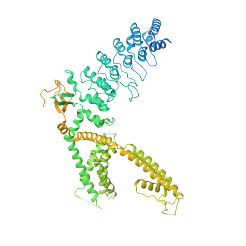
Structural basis of TRPV3 inhibition by an antagonist.

Fan, J.Hu, L.Yue, Z.Liao, D.Guo, F.Ke, H.Jiang, D.Yang, Y.Lei, X.

(2023) Nat Chem Biol 19: 81-90

  • DOI: https://doi.org/10.1038/s41589-022-01166-5
  • Primary Citation of Related Structures:  
    7XJ0, 7XJ1, 7XJ2, 7XJ3

  • PubMed Abstract: 

    The TRPV3 channel plays vital roles in skin physiology. Dysfunction of TRPV3 causes skin diseases, including Olmsted syndrome. However, the lack of potent and selective inhibitors impedes the validation of TRPV3 as a therapeutic target. In this study, we identified Trpvicin as a potent and subtype-selective inhibitor of TRPV3. Trpvicin exhibits pharmacological potential in the inhibition of itch and hair loss in mouse models. Cryogenic electron microscopy structures of TRPV3 and the pathogenic G573S mutant complexed with Trpvicin reveal detailed ligand-binding sites, suggesting that Trpvicin inhibits the TRPV3 channel by stabilizing it in a closed state. Our G573S mutant structures demonstrate that the mutation causes a dilated pore, generating constitutive opening activity. Trpvicin accesses additional binding sites inside the central cavity of the G573S mutant to remodel the channel symmetry and block the channel. Together, our results provide mechanistic insights into the inhibition of TRPV3 by Trpvicin and support TRPV3-related drug development.


  • Organizational Affiliation

    Beijing National Laboratory for Molecular Sciences, Key Laboratory of Bioorganic Chemistry and Molecular Engineering of Ministry of Education, Department of Chemical Biology, College of Chemistry and Molecular Engineering, Synthetic and Functional Biomolecules Center and Peking-Tsinghua Center for Life Sciences, Peking University, Beijing, China.


Macromolecules
Find similar proteins by:  (by identity cutoff)  |  3D Structure
Entity ID: 1
MoleculeChains Sequence LengthOrganismDetailsImage
Fusion protein of Transient receptor potential cation channel subfamily V member 3 and 3C-GFPA [auth B],
B [auth C],
C [auth A],
D
1,061Homo sapiensMutation(s): 0 
Gene Names: TRPV3
Membrane Entity: Yes 
UniProt & NIH Common Fund Data Resources
Find proteins for Q8NET8 (Homo sapiens)
Explore Q8NET8 
Go to UniProtKB:  Q8NET8
PHAROS:  Q8NET8
GTEx:  ENSG00000167723 
Entity Groups  
Sequence Clusters30% Identity50% Identity70% Identity90% Identity95% Identity100% Identity
UniProt GroupQ8NET8
Sequence Annotations
Expand
  • Reference Sequence
Small Molecules
Ligands 2 Unique
IDChains Name / Formula / InChI Key2D Diagram3D Interactions
6OU
Query on 6OU

Download Ideal Coordinates CCD File 
E [auth B]
H [auth B]
I [auth B]
K [auth C]
L [auth C]
E [auth B],
H [auth B],
I [auth B],
K [auth C],
L [auth C],
M [auth C],
R [auth A],
S [auth A],
V [auth D],
X [auth D]
[(2~{R})-1-[2-azanylethoxy(oxidanyl)phosphoryl]oxy-3-hexadecanoyloxy-propan-2-yl] (~{Z})-octadec-9-enoate
C39 H76 N O8 P
FHQVHHIBKUMWTI-OTMQOFQLSA-N
EQK (Subject of Investigation/LOI)
Query on EQK

Download Ideal Coordinates CCD File 
F [auth B]
G [auth B]
J [auth C]
N [auth C]
O [auth C]
F [auth B],
G [auth B],
J [auth C],
N [auth C],
O [auth C],
P [auth A],
Q [auth A],
T [auth A],
U [auth D],
W [auth D]
N-[5-[2-(2-cyanopropan-2-yl)pyridin-4-yl]-4-(trifluoromethyl)-1,3-thiazol-2-yl]-4,6-dimethoxy-pyrimidine-5-carboxamide
C20 H17 F3 N6 O3 S
CPIDEBBXBVMQOC-UHFFFAOYSA-N
Experimental Data & Validation

Experimental Data

  • Method: ELECTRON MICROSCOPY
  • Resolution: 2.93 Å
  • Aggregation State: PARTICLE 
  • Reconstruction Method: SINGLE PARTICLE 

Structure Validation

View Full Validation Report


Entry History & Funding Information

Deposition Data


Funding OrganizationLocationGrant Number
National Natural Science Foundation of China (NSFC)China21625201

Revision History  (Full details and data files)

  • Version 1.0: 2022-11-09
    Type: Initial release
  • Version 1.1: 2022-11-16
    Changes: Database references
  • Version 1.2: 2023-01-11
    Changes: Database references