


Crystal structure of SAV2152 from MRSA
Park, H.J., Seok, S.H., Kim, J.H.To be published.
Experimental Data Snapshot
wwPDB Validation 3D Report Full Report

Entity ID: 1 | |||||
|---|---|---|---|---|---|
| Molecule | Chains | Sequence Length | Organism | Details | Image |
| Phosphatase, SAV2152 | 231 | Staphylococcus aureus subsp. aureus Mu50 | Mutation(s): 0 Gene Names: SAV2152 | 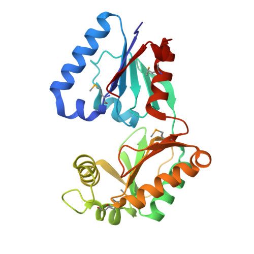 | |
UniProt | |||||
Find proteins for A0A0H3JWK0 (Staphylococcus aureus (strain Mu50 / ATCC 700699)) Explore A0A0H3JWK0 Go to UniProtKB: A0A0H3JWK0 | |||||
Entity Groups | |||||
| Sequence Clusters | 30% Identity50% Identity70% Identity90% Identity95% Identity100% Identity | ||||
| UniProt Group | A0A0H3JWK0 | ||||
Sequence AnnotationsExpand | |||||
Reference Sequence | |||||
| Modified Residues 1 Unique | |||||
|---|---|---|---|---|---|
| ID | Chains | Type | Formula | 2D Diagram | Parent |
| MSE Query on MSE | A | L-PEPTIDE LINKING | C5 H11 N O2 Se |  | MET |
| Length ( Å ) | Angle ( ˚ ) |
|---|---|
| a = 53.61 | α = 90 |
| b = 63.06 | β = 90 |
| c = 92.73 | γ = 90 |
| Software Name | Purpose |
|---|---|
| PHENIX | refinement |
| PHENIX | phasing |
| HKL-2000 | data scaling |
| PHENIX | model building |
| Funding Organization | Location | Grant Number |
|---|---|---|
| Not funded | -- |


