7XEO | pdb_00007xeo

MbetaCD treated state of mTRPV2


Experimental Data Snapshot

  • Method: ELECTRON MICROSCOPY
  • Resolution: 2.89 Å
  • Aggregation State: PARTICLE 
  • Reconstruction Method: SINGLE PARTICLE 

wwPDB Validation 3D Report Full Report

Validation slider image for 7XEO

This is version 1.3 of the entry. See complete history

Literature

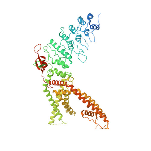
Structural mechanisms of TRPV2 modulation by endogenous and exogenous ligands.

Su, N.Zhen, W.Zhang, H.Xu, L.Jin, Y.Chen, X.Zhao, C.Wang, Q.Wang, X.Li, S.Wen, H.Yang, W.Guo, J.Yang, F.

(2023) Nat Chem Biol 19: 72-80

  • DOI: https://doi.org/10.1038/s41589-022-01139-8
  • Primary Citation Related Structures: 
    7XEM, 7XEO, 7XER, 7XEU, 7XEV, 7XEW, 7YEP

  • PubMed Abstract: 

    The transient receptor potential vanilloid 2 (TRPV2) ion channel is a polymodal receptor widely involved in many physiological and pathological processes. Despite many TRPV2 modulators being identified, whether and how TRPV2 is regulated by endogenous lipids remains elusive. Here, we report an endogenous cholesterol molecule inside the vanilloid binding pocket (VBP) of TRPV2, with a 'head down, tail up' configuration, resolved at 3.2 Å using cryo-EM. Cholesterol binding antagonizes ligand activation of TRPV2, which is removed from VBP by methyl-β-cyclodextrin (MβCD) as resolved at 2.9 Å. We also observed that estradiol (E2) potentiated TRPV2 activation by 2-aminoethoxydiphenyl borate (2-APB), a classic tool compound for TRP channels. Our cryo-EM structures (resolved at 2.8-3.3 Å) further suggest how E2 disturbed cholesterol binding and how 2-APB bound within the VBP with E2 or without both E2 and endogenous cholesterol, respectively. Therefore, our study has established the structural basis for ligand recognition of the inhibitory endogenous cholesterol and excitatory exogenous 2-APB in TRPV2.


  • Organizational Affiliation
    • Department of Biophysics, and Kidney Disease Center of the First Affiliated Hospital, Zhejiang University School of Medicine, Hangzhou, China.

Macromolecule Content 

  • Total Structure Weight: 344.23 kDa 
  • Atom Count: 19,980 
  • Modeled Residue Count: 2,456 
  • Deposited Residue Count: 3,024 
  • Unique protein chains: 1

Macromolecules

Find similar proteins by:|  3D Structure
Entity ID: 1
MoleculeChains  Sequence LengthOrganismDetailsImage
Transient receptor potential cation channel subfamily V member 2
A, B, C, D
756Mus musculusMutation(s): 0 
Gene Names: Trpv2Grc
Membrane Entity: Yes 
UniProt
Find proteins for Q9WTR1 (Mus musculus)
Explore Q9WTR1 
Go to UniProtKB:  Q9WTR1
Entity Groups
Sequence Clusters30% Identity50% Identity70% Identity90% Identity95% Identity100% Identity
UniProt GroupQ9WTR1
Sequence Annotations
Expand
Reference Sequence

Experimental Data & Validation

Experimental Data

  • Method: ELECTRON MICROSCOPY
  • Resolution: 2.89 Å
  • Aggregation State: PARTICLE 
  • Reconstruction Method: SINGLE PARTICLE 
EM Software:
TaskSoftware PackageVersion
MODEL REFINEMENTPHENIX

Structure Validation

View Full Validation Report


Entry History 

& Funding Information

Deposition Data

  • Released Date: 2022-08-17 
  • Deposition Author(s): Su, N.

Funding OrganizationLocationGrant Number
National Natural Science Foundation of China (NSFC)China32122040

Revision History  (Full details and data files)

  • Version 1.0: 2022-08-17
    Type: Initial release
  • Version 1.1: 2023-03-01
    Changes: Database references, Structure summary
  • Version 1.2: 2024-06-26
    Changes: Data collection
  • Version 1.3: 2025-06-25
    Changes: Data collection, Structure summary