Chlorine Atoms of an Aripiprazole Molecule Control the Geometry and Motion of Aripiprazole and Deschloro-aripiprazole in Subdomain IIIA of Human Serum Albumin.
Kawai, A., Kobashigawa, Y., Hirata, K., Morioka, H., Imoto, S., Nishi, K., Chuang, V.T.G., Yamasaki, K., Otagiri, M.(2022) ACS Omega 7: 29944-29951
- PubMed: 36061730 Search on PubMedSearch on PubMed Central
- DOI: https://doi.org/10.1021/acsomega.2c02929
- Primary Citation Related Structures:
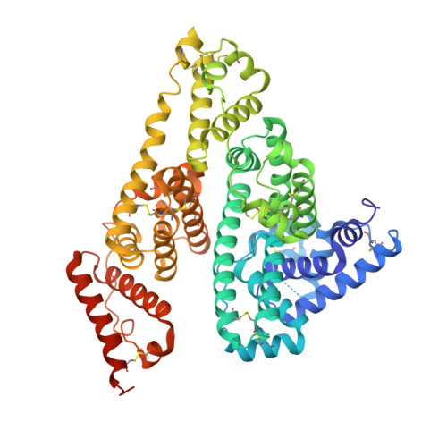
7X7X - PubMed Abstract:
Aripiprazole (ARP), an antipsychotic drug, binds more strongly to human serum albumin (HSA) than the other ARP derivatives. In addition, the signs for the extrinsic Cotton effects for HSA complexed with ARP or deschloro-ARP are reversed. In this study, we report on a structural-chemical approach using circular dichroism (CD) spectroscopic analysis, X-ray crystallographic analysis, and molecular dynamics simulations. The objective was to examine the relationship between the induced CD spectra and the structural features of the HSA complexes with ARP or deschloro-ARP. The intensity of the induced CD spectra of the HSA complexes with ARP or deschloro-ARP was reduced with increasing temperature. We determined the crystal structure of the HSA complexed with deschloro-ARP in this study and compared it to HSA complexed with ARP that we reported previously. The comparison of these structures revealed that both ARP and deschloro-ARP were bound at the site II pocket in HSA and that the orientation of the molecules was nearly identical. Molecular dynamics simulations indicated that the molecular motions of ARP and deschloro-ARP within the site II pocket were different from one another and the proportion of stacking interaction formations of Tyr411 with the dihydroquinoline rings of ARP and deschloro-ARP was also different. These findings indicate that the induced CD spectra are related to the molecular motions and dynamic interactions of ARP and deschloro-ARP in HSA and may help to understand the molecular recognition and motion that occurs within the binding site for the other HSA ligands more clearly.
- Fujita Health University School of Medicine, 1-98 Dengakugakubo, Kutsukake-cho, Toyoake, Aichi 470-1192, Japan.
Organizational Affiliation: