


The crystal structure of human Calpain-1 protease core in complex with 14a
Zhao, Y., Zhao, J., Shao, M., Yang, H., Rao, Z.To be published.
Experimental Data Snapshot
Starting Model: experimental
View more details
Entity ID: 1 | |||||
|---|---|---|---|---|---|
| Molecule | Chains | Sequence Length | Organism | Details | Image |
| Calpain-1 catalytic subunit | 714 | Homo sapiens | Mutation(s): 0 Gene Names: CAPN1, CANPL1, PIG30 EC: 3.4.22.52 | 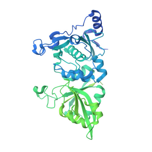 | |
UniProt & NIH Common Fund Data Resources | |||||
PHAROS: P07384 GTEx: ENSG00000014216 | |||||
Entity Groups | |||||
| Sequence Clusters | 30% Identity50% Identity70% Identity90% Identity95% Identity100% Identity | ||||
| UniProt Group | P07384 | ||||
Sequence AnnotationsExpand | |||||
Reference Sequence | |||||
| Ligands 3 Unique | |||||
|---|---|---|---|---|---|
| ID | Chains | Name / Formula / InChI Key | 2D Diagram | 3D Interactions | |
| 89K (Subject of Investigation/LOI) Download:Ideal Coordinates CCD File | B [auth A] | N-[(2S)-3-cyclohexyl-1-oxidanylidene-1-[[(2S,3S)-3-oxidanyl-4-oxidanylidene-1-[(3S)-2-oxidanylidenepiperidin-3-yl]-4-[(phenylmethyl)amino]butan-2-yl]amino]propan-2-yl]-1-benzofuran-2-carboxamide C34 H42 N4 O6 QBWBVQMSMXVSSF-YRUYCOJQSA-N |  | ||
| CA Download:Ideal Coordinates CCD File | D [auth A], E [auth A] | CALCIUM ION Ca BHPQYMZQTOCNFJ-UHFFFAOYSA-N |  | ||
| H2S Download:Ideal Coordinates CCD File | C [auth A] | HYDROSULFURIC ACID H2 S RWSOTUBLDIXVET-UHFFFAOYSA-N |  | ||
| Length ( Å ) | Angle ( ˚ ) |
|---|---|
| a = 50.203 | α = 90 |
| b = 64.07 | β = 90 |
| c = 99.576 | γ = 90 |
| Software Name | Purpose |
|---|---|
| PHENIX | refinement |
| XSCALE | data scaling |
| PDB_EXTRACT | data extraction |
| XDS | data reduction |
| PHASER | phasing |
| Funding Organization | Location | Grant Number |
|---|---|---|
| National Natural Science Foundation of China (NSFC) | China | 813300237 |


