7X4H

Crystal structure of CK2a1 complexed with AG1112


Experimental Data Snapshot

  • Method: X-RAY DIFFRACTION
  • Resolution: 1.77 Å
  • R-Value Free: 0.208 
  • R-Value Work: 0.169 
  • R-Value Observed: 0.171 

wwPDB Validation   3D Report Full Report


Ligand Structure Quality Assessment 


This is version 1.1 of the entry. See complete history


Literature

Bivalent binding mode of an amino-pyrazole inhibitor indicates the potentials for CK2 alpha 1-selective inhibitors.

Ikeda, A.Tsuyuguchi, M.Kitagawa, D.Sawa, M.Nakamura, S.Nakanishi, I.Kinoshita, T.

(2022) Biochem Biophys Res Commun 630: 30-35

  • DOI: https://doi.org/10.1016/j.bbrc.2022.09.040
  • Primary Citation of Related Structures:  
    7X4H, 7XYH

  • PubMed Abstract: 

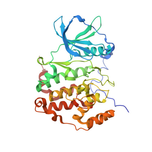
    Casein kinase 2 (CK2) is a vital protein kinase that consists of two catalytic subunits (CK2α1 and/or CK2α2) and two regulatory subunits (CK2β). CK2α1 is a drug target for nephritis and cancers, while CK2α2 is a serious off-target because its inhibition causes testicular toxicity. High similarity between the isozymes CK2α1 and CK2α2 make it difficult to design CK2α1-specific inhibitors. Herein, the crystal structures of CK2α1 and CK2α2 complexed with a 3-amino-pyrazole inhibitor revealed the remarkable differences in the protein-inhibitor interaction modes. This inhibitor bound to the ATP binding sites of both isozymes in apparently distinct orientations. In addition, another molecule of this inhibitor bound to CK2α1, but not to CK2α2, at the CK2β protein-protein interface. Binding energy calculations and biochemical experiments suggested that this inhibitor possesses the conventional ATP-competitive characteristics with moderate allosteric function in a molecular glue mechanism. These results will assist the potential design of potent and selective CK2α1 inhibitors.


  • Organizational Affiliation

    Graduate School of Science, Osaka Metropolitan University, Sakai, 599-8531, Japan.


Macromolecules
Find similar proteins by:  (by identity cutoff)  |  3D Structure
Entity ID: 1
MoleculeChains Sequence LengthOrganismDetailsImage
Casein Kinase 2 subunit alpha340Homo sapiensMutation(s): 0 
EC: 2.7.11.1
UniProt & NIH Common Fund Data Resources
Find proteins for P68400 (Homo sapiens)
Explore P68400 
Go to UniProtKB:  P68400
PHAROS:  P68400
GTEx:  ENSG00000101266 
Entity Groups  
Sequence Clusters30% Identity50% Identity70% Identity90% Identity95% Identity100% Identity
UniProt GroupP68400
Sequence Annotations
Expand
  • Reference Sequence
Small Molecules
Ligands 1 Unique
IDChains Name / Formula / InChI Key2D Diagram3D Interactions
8BH
Query on 8BH

Download Ideal Coordinates CCD File 
B [auth A],
C [auth A]
5-azanyl-3-[(~{Z})-1-cyano-2-(1~{H}-indol-3-yl)ethenyl]-1~{H}-pyrazole-4-carbonitrile
C15 H10 N6
OJMXHLJIMZMFGX-WEVVVXLNSA-N
Experimental Data & Validation

Experimental Data

  • Method: X-RAY DIFFRACTION
  • Resolution: 1.77 Å
  • R-Value Free: 0.208 
  • R-Value Work: 0.169 
  • R-Value Observed: 0.171 
  • Space Group: P 21 21 21
Unit Cell:
Length ( Å )Angle ( ˚ )
a = 48.025α = 90
b = 79.377β = 90
c = 82.26γ = 90
Software Package:
Software NamePurpose
PHENIXrefinement
HKL-2000data reduction
HKL-2000data scaling
PHENIXphasing

Structure Validation

View Full Validation Report


Ligand Structure Quality Assessment 


Entry History & Funding Information

Deposition Data


Funding OrganizationLocationGrant Number
Not funded--

Revision History  (Full details and data files)

  • Version 1.0: 2023-01-18
    Type: Initial release
  • Version 1.1: 2023-11-29
    Changes: Data collection, Refinement description