7X2E

Structure of USH1C PDZ2 and coiled-coil in complex with CDHR2 C-terminal tail


Experimental Data Snapshot

  • Method: X-RAY DIFFRACTION
  • Resolution: 1.85 Å
  • R-Value Free: 0.216 
  • R-Value Work: 0.181 
  • R-Value Observed: 0.182 

wwPDB Validation   3D Report Full Report


This is version 2.0 of the entry. See complete history


Literature

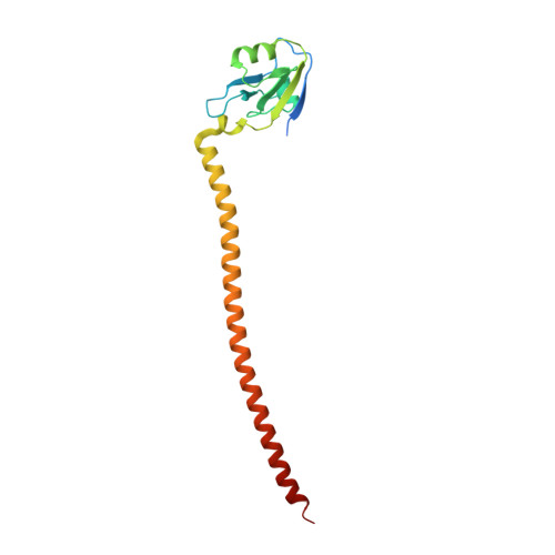
Structure of the Harmonin PDZ2 and coiled-coil domains in a complex with CDHR2 tail and its implications.

Yan, W.Chen, G.Li, J.

(2022) FASEB J 36: e22425-e22425

  • DOI: https://doi.org/10.1096/fj.202200403RR
  • Primary Citation of Related Structures:  
    7X2E

  • PubMed Abstract: 

    Harmonin is a protein containing multiple PDZ domains and is required for the development and maintenance of hair cell stereocilia and brush border microvilli. Mutations in the USH1C gene can cause Usher syndrome type 1C, a severe inheritable disease characterized by the loss of hearing and vision. Here, by solving the high-resolution crystal structure of Harmonin PDZ2 and coiled-coil domains in a complex with the tail of cadherin-related family member 2, we demonstrated that mutations located in the Harmonin PDZ2 domain and found in patients could affect its stability, and thus, the target binding capability. The structure also implies that the coiled-coil domain could form antiparallel dimers under high concentrations, possibly when Harmonin underwent liquid-liquid phase separation in the upper tip-link density in hair cell stereocilia or microvilli of enterocytes of the intestinal epithelium. The crystal structure, together with the biochemical analysis, provided mechanistic implications for Harmonin mutations causing Usher syndrome, non-syndromic deafness, or enteropathy.


  • Organizational Affiliation

    School of Biology and Biological Engineering, South China University of Technology, Guangzhou, China.


Macromolecules
Find similar proteins by:  (by identity cutoff)  |  3D Structure
Entity ID: 1
MoleculeChains Sequence LengthOrganismDetailsImage
Harmonin178Homo sapiensMutation(s): 0 
Gene Names: USH1C
UniProt & NIH Common Fund Data Resources
Find proteins for Q9Y6N9 (Homo sapiens)
Explore Q9Y6N9 
Go to UniProtKB:  Q9Y6N9
PHAROS:  Q9Y6N9
GTEx:  ENSG00000006611 
Entity Groups  
Sequence Clusters30% Identity50% Identity70% Identity90% Identity95% Identity100% Identity
UniProt GroupQ9Y6N9
Sequence Annotations
Expand
  • Reference Sequence

Find similar proteins by:  Sequence   |   3D Structure  

Entity ID: 2
MoleculeChains Sequence LengthOrganismDetailsImage
Cadherin-related family member 218Mus musculusMutation(s): 0 
UniProt
Find proteins for E9Q7P9 (Mus musculus)
Explore E9Q7P9 
Go to UniProtKB:  E9Q7P9
Entity Groups  
Sequence Clusters30% Identity50% Identity70% Identity90% Identity95% Identity100% Identity
UniProt GroupE9Q7P9
Sequence Annotations
Expand
  • Reference Sequence
Experimental Data & Validation

Experimental Data

  • Method: X-RAY DIFFRACTION
  • Resolution: 1.85 Å
  • R-Value Free: 0.216 
  • R-Value Work: 0.181 
  • R-Value Observed: 0.182 
  • Space Group: C 1 2 1
Unit Cell:
Length ( Å )Angle ( ˚ )
a = 54.516α = 90
b = 50.681β = 107.77
c = 72.847γ = 90
Software Package:
Software NamePurpose
DENZOdata reduction
SCALEPACKdata scaling
PHENIXrefinement
PDB_EXTRACTdata extraction
PHASERphasing

Structure Validation

View Full Validation Report


Entry History & Funding Information

Deposition Data


Funding OrganizationLocationGrant Number
National Natural Science Foundation of China (NSFC)China--

Revision History  (Full details and data files)

  • Version 1.0: 2022-07-06
    Type: Initial release
  • Version 2.0: 2023-11-29
    Changes: Atomic model, Data collection, Derived calculations, Refinement description